
Mitral valve fixation device removal devices and methods

a technology of fixation device and mitral valve, which is applied in the field of medical methods, devices and systems, can solve the problems that the mitral valve may not be suitable candidates for fixation device, may still exist or could arise with the function of the mitral valve or the heart generally, and may not be suitable candidates for the mitral valv

Active Publication Date: 2017-02-21
EVALVE
View PDF55 Cites 20 Cited by
  • Summary
  • Abstract
  • Description
  • Claims
  • Application Information

AI Technical Summary

Benefits of technology

Enables safe and less invasive removal or disabling of mitral valve fixation devices, reducing morbidity and allowing for subsequent cardiac procedures without the need for open-heart surgery, even in patients with complex cardiac anatomy.

Problems solved by technology

Sometimes after such a device is installed in the heart, problems may still exist or could arise with the function of the mitral valve or with the heart generally.
However, less invasive methods would be preferable, because, for example, persons with a mitral valve fixation device may not be suitable candidates for an invasive surgery.

Method used


Image

  • Mitral valve fixation device removal devices and methods
  • Mitral valve fixation device removal devices and methods
  • Mitral valve fixation device removal devices and methods

Examples


Embodiment Construction

I. Introduction

A. Cardiac Physiology

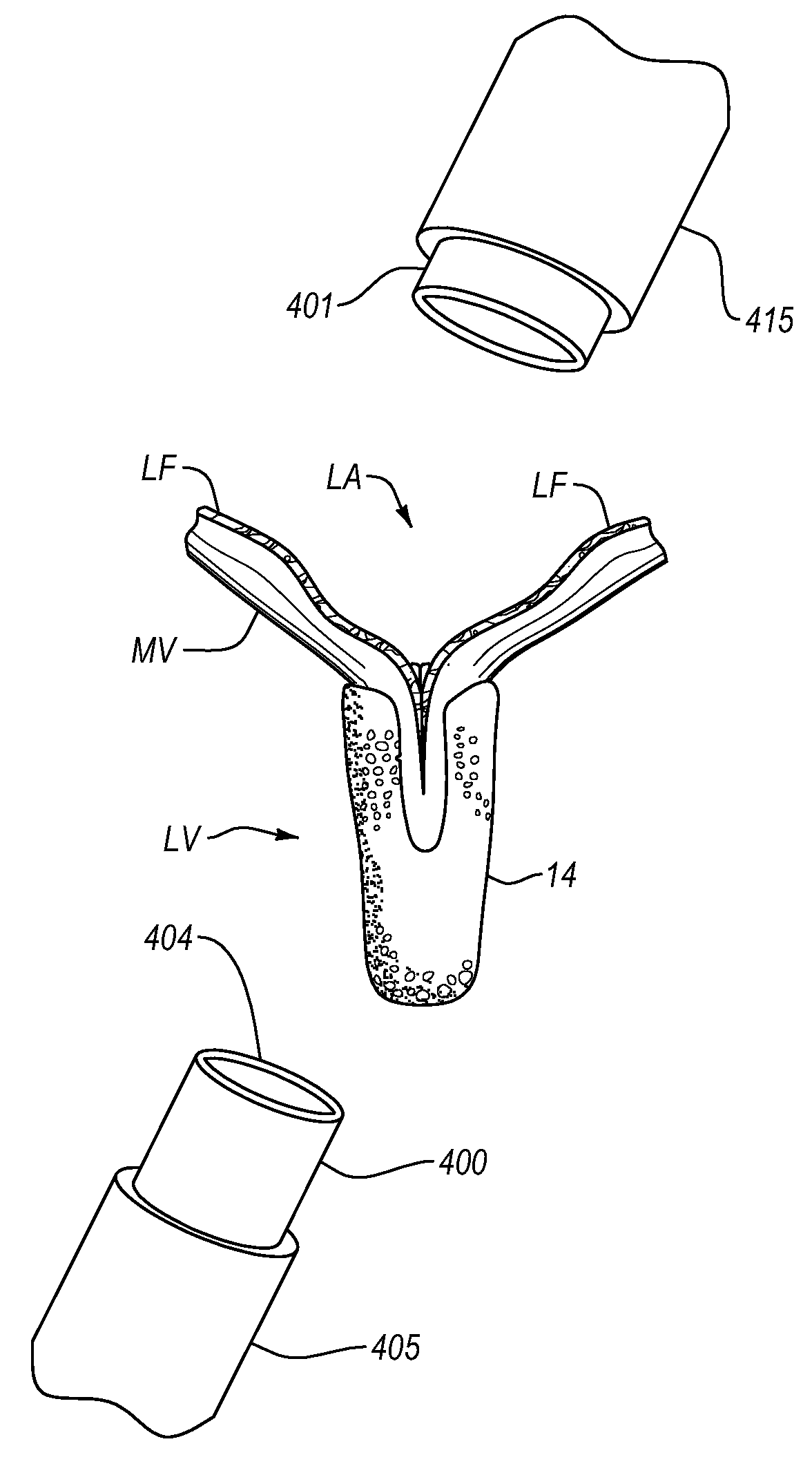
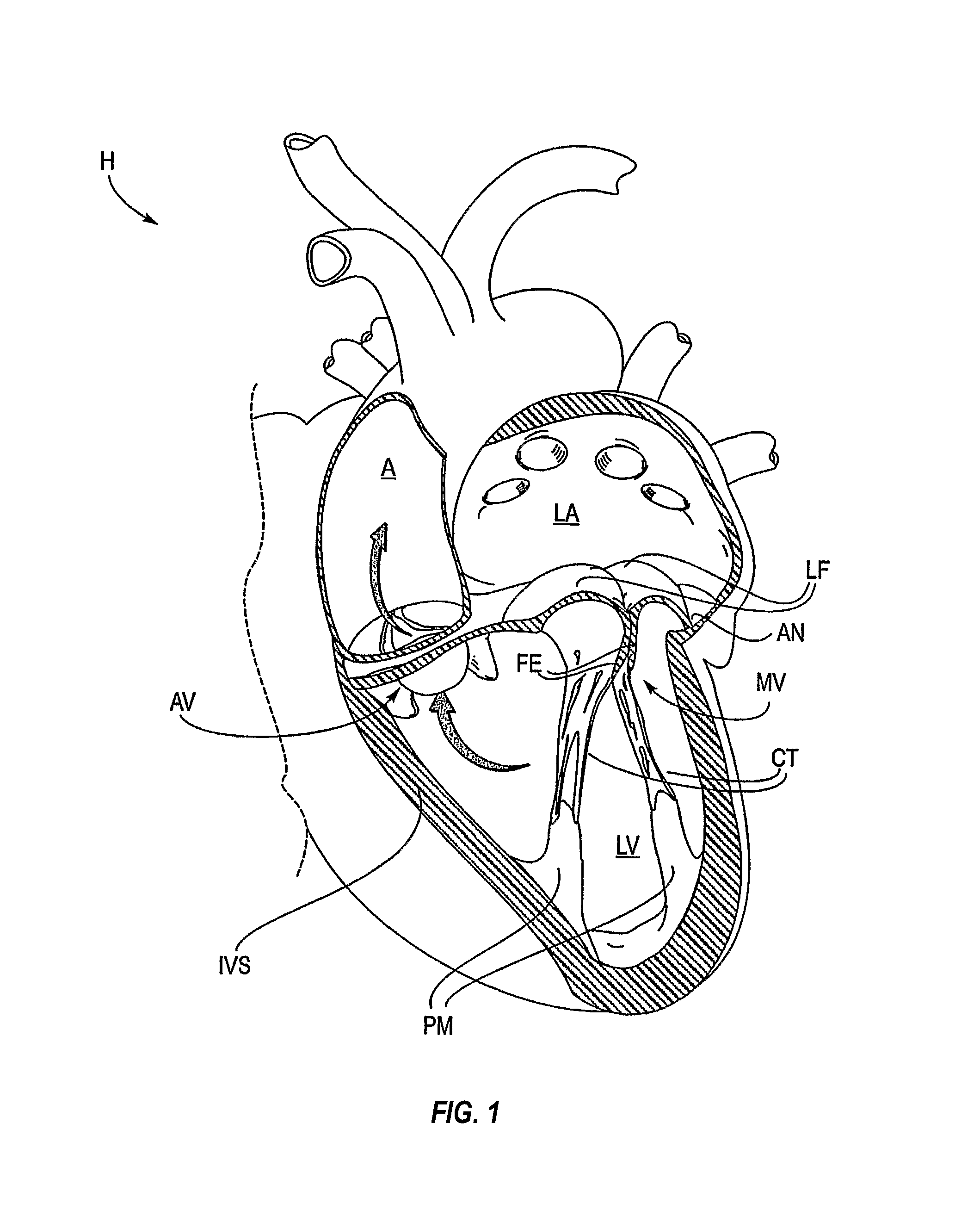
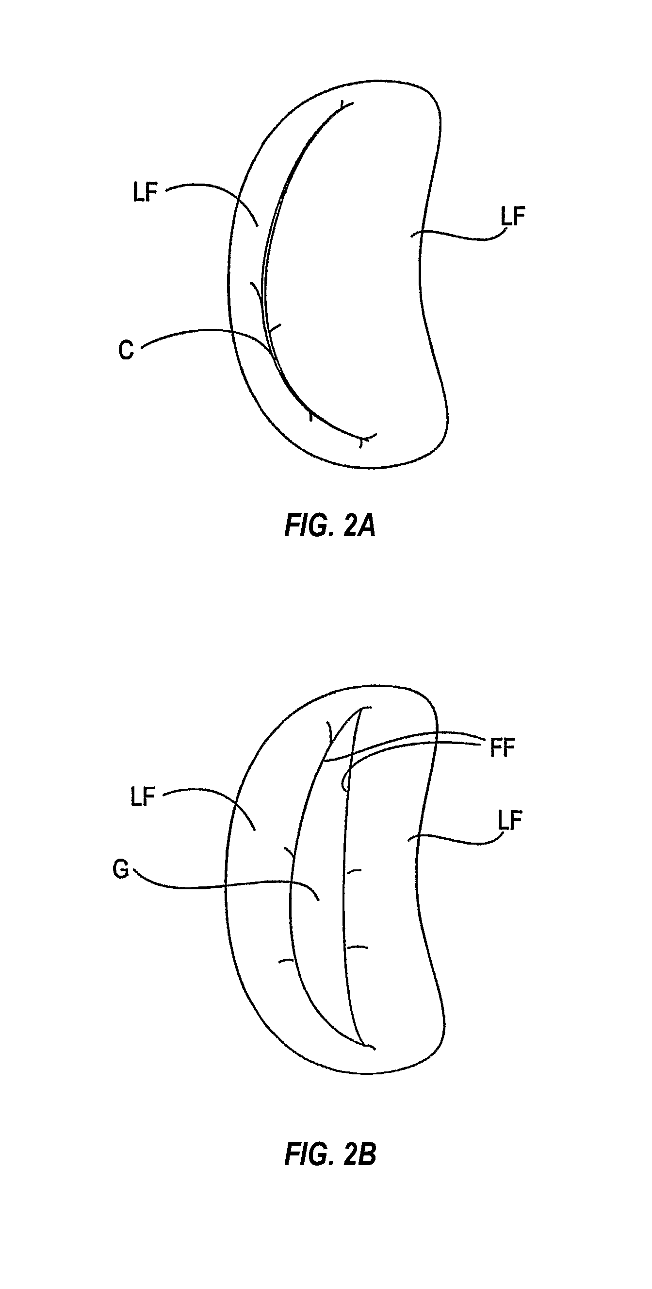
[0059]The left ventricle (LV) of a normal heart H in systole is illustrated in FIG. 1. The left ventricle (LV) is contracting and blood flows outwardly through the tricuspid (aortic) valve (AV) in the direction of the arrows. Back flow of blood or “regurgitation” through the mitral valve (MV) is prevented since the mitral valve is configured as a “check valve” which prevents back flow when pressure in the left ventricle is higher than that in the left atrium (LA). The mitral valve (MV) comprises a pair of leaflets having free edges (FE) which meet evenly to close, as illustrated in FIG. 1. The opposite ends of the leaflets (LF) are attached to the surrounding heart structure along an annular region referred to as the annulus (AN). The free edges (FE) of the leaflets (LF) are secured to the lower portions of the left ventricle LV through chordae tendinae (CT) (referred to hereinafter as the chordae) which include a plurality of branching tendons se...


PUM


Abstract

Procedures may be performed on the heart after the installation of a mitral valve fixation device. In order to prepare the heart for such procedures, the fixation device may be removed or disabled in minimally invasive ways (e.g., through an endovascular procedure), without requiring open access to the heart. The fixation device may be partitioned so that one portion may remain attached to each leaflet of the mitral valve. In another example, the leaflets may be cut along the edges of the distal element(s) of the fixation device, so as to cut the fixation device from the leaflet(s). Systems and devices for performing such procedures endovascularly are disclosed. Fixation devices with improved access to a release harness are also disclosed.

Description

CROSS-REFERENCE TO RELATED APPLICATIONS[0001]NABACKGROUND[0002]The present invention relates generally to medical methods, devices, and systems. In particular, the present invention relates to methods, devices, and systems for the endovascular, percutaneous or minimally invasive surgical treatment of bodily tissues, such as tissue approximation or valve repair. More particularly, the present invention relates to repair of valves of the heart and venous valves, and devices and methods for removing or disabling mitral valve repair components through minimally invasive procedures.[0003]Surgical repair of bodily tissues often involves tissue approximation and fastening of such tissues in the approximated arrangement. When repairing valves, tissue approximation includes coapting the leaflets of the valves in a therapeutic arrangement which may then be maintained by fastening or fixing the leaflets. Such coaptation can be used to treat regurgitation which most commonly occurs in the mitra...

Claims


Application Information

Patent Timeline
Patent Type & Authority Patents(United States)
IPC IPC(8): A61F2/24A61B17/068A61B17/064A61B17/22A61B17/00A61B17/3205
CPCA61B17/0682A61F2/2466A61F2/2463A61B17/0644A61B17/32053A61B2017/00243A61B2017/00783A61B2017/00867A61B2017/22097A61F2/246A61B17/0467
Inventor BASUDE, RAGHUVEERDELL, KENTKABE, ARUNDHATIKRISHNAMURTHY, GAURAVWEI, MICHAEL F.
Owner EVALVE
Learn More