Excised specimen imaging using a combined PET and micro CT scanner

a combined pet and micro ct technology, applied in the direction of tomography, patient positioning for diagnostics, instruments, etc., can solve the problems of impeded accurate identification of which margins of specimens, insufficient information for accurate assesment of whether sufficient tissue was removed, and several days to obtain

Active Publication Date: 2017-01-31
RGT UNIV OF CALIFORNIA
View PDF26 Cites 11 Cited by
  • Summary
  • Abstract
  • Description
  • Claims
  • Application Information

AI Technical Summary

Problems solved by technology

However, as both wires and clips can move during surgery, detection of these lesion markers provides insufficient information for accurate assesment of whether sufficient tissue was removed.
Further, the two-dimensional nature of X-ray radiography and the compression required impede accurate identification of which margins of the specimen are clear of cancerous tissue.
Histopathology results often require several days to obtain due to the time required for specimen logging, grossing, preservation, paraffinization, cutting, staining, and microscopic review.
The need for breast cancer patients to undergo a second surgery (and, potentially, additional subsequent surgeries) creates significant emotional and financial stress.

Method used


Image

  • Excised specimen imaging using a combined PET and micro CT scanner
  • Excised specimen imaging using a combined PET and micro CT scanner
  • Excised specimen imaging using a combined PET and micro CT scanner

Examples


Embodiment Construction

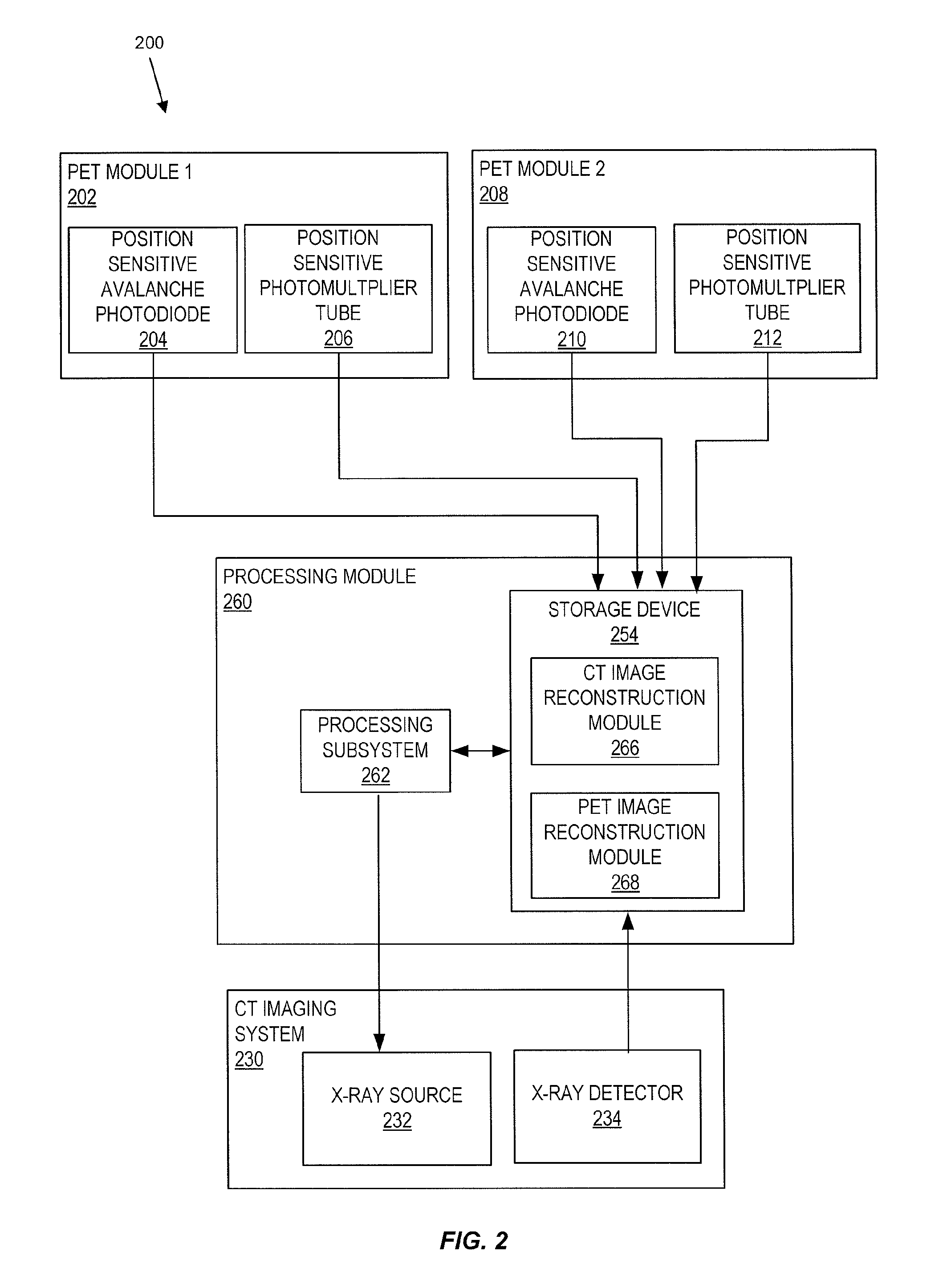
[0015]Embodiments of the present invention relate to imaging a tissue specimen using a combined positron emission tomography (PET) and micro computed tomography (micro CT) scanner. A PET imaging system and a micro CT imaging system can be mounted to a gantry. Typically, the gantry is sufficiently compact to be mounted on a cart that can be used in used in or adjacent to an operating room.

[0016]An image constructued based on data acquired from the micro CT imaging system can show calcificaitons and lesion markers in the tissue specimen. Lesion markers such as clips and / or guide wires inserted into the body prior to surgery can help a surgeon locate a lesion (e.g., tumor) during surgery. If lesion markers that were inserted are not present in a tissue sample removed, this may be an indicator that the surgeon will need to remove additional tissue. The presence of calcifications or cancerous tissue close to the surface (i.e. in the margin) of the specimen are also potential indicators t...


PUM

PropertyMeasurementUnit
depthaaaaaaaaaa
depthaaaaaaaaaa
depthaaaaaaaaaa

Abstract

Embodiments of the present invention provide methods and apparatus for imaging a tissue specimen excised during surgery with a combined positron emission tomography (PET) and micro computed tomography (micro CT) scanner. The specimen is scanned with a CT imaging system of the combined PET and micro CT scanner. The specimen is also scanned with a PET imaging system of the combined PET and micro CT scanner. A PET image is constructed based on data acquired by the PET imaging system. A micro CT image is constructed based on data acquired by the micro CT imaging system. The micro CT image includes at least one visualization of a lesion marker.

Description

CROSS-REFERENCES TO RELATED APPLICATIONS[0001]The present application claims priority to U.S. Patent Application No. 61 / 495,340, filed Jun. 9, 2011, which is hereby incorporated by reference, as if set forth in full in this document, for all purposes.STATEMENT AS TO RIGHTS TO INVENTIONS MADE UNDER FEDERALLY SPONSORED RESEARCH AND DEVELOPMENT[0002]This invention was made with Government support under Grant Nos. CA 129561 & EB002138, awarded by the National Institutes of Health. The Government has certain rights in this invention.BACKGROUND[0003]Surgical treatments including mastectomy and lumpectomy are applied to remove tumors from patients having breast cancer. Optimal outcomes require tumorous tissue to be surgically removed as completely as possible. To aid in removal of tumors, a radiologist may insert a clip in a breast duing biopsy to mark the tumor. A guide wire or multiple guide wires may be inserted into the breast to help the surgeon locate the targeted tumor during surger...

Claims


Application Information

Patent Timeline
Patent Type & Authority Patents(United States)
IPC IPC(8): G01N23/04A61B6/03G01T1/161A61B6/00A61B6/04
CPCG01N23/046A61B6/032A61B6/037A61B6/4258A61B6/508A61B6/5235G01T1/161A61B6/0407G01N2223/108G01N2223/1016G01N2223/419G01N2223/6126
Inventor BADAWI, RAMSEY D.YANG, KAI
Owner RGT UNIV OF CALIFORNIA
Why Eureka
Try Eureka