Acellular vascular products
a technology of vascular products and a cell, applied in the field of acellular vascular products, can solve the problems of rejection of xenografts, inability to perform xenotransplantation trials completely, and inability to achieve complete success in treatment, etc., and achieve the effect of successful treatmen
- Summary
- Abstract
- Description
- Claims
- Application Information
AI Technical Summary
Benefits of technology
Problems solved by technology
Method used
Image
Examples
example 1
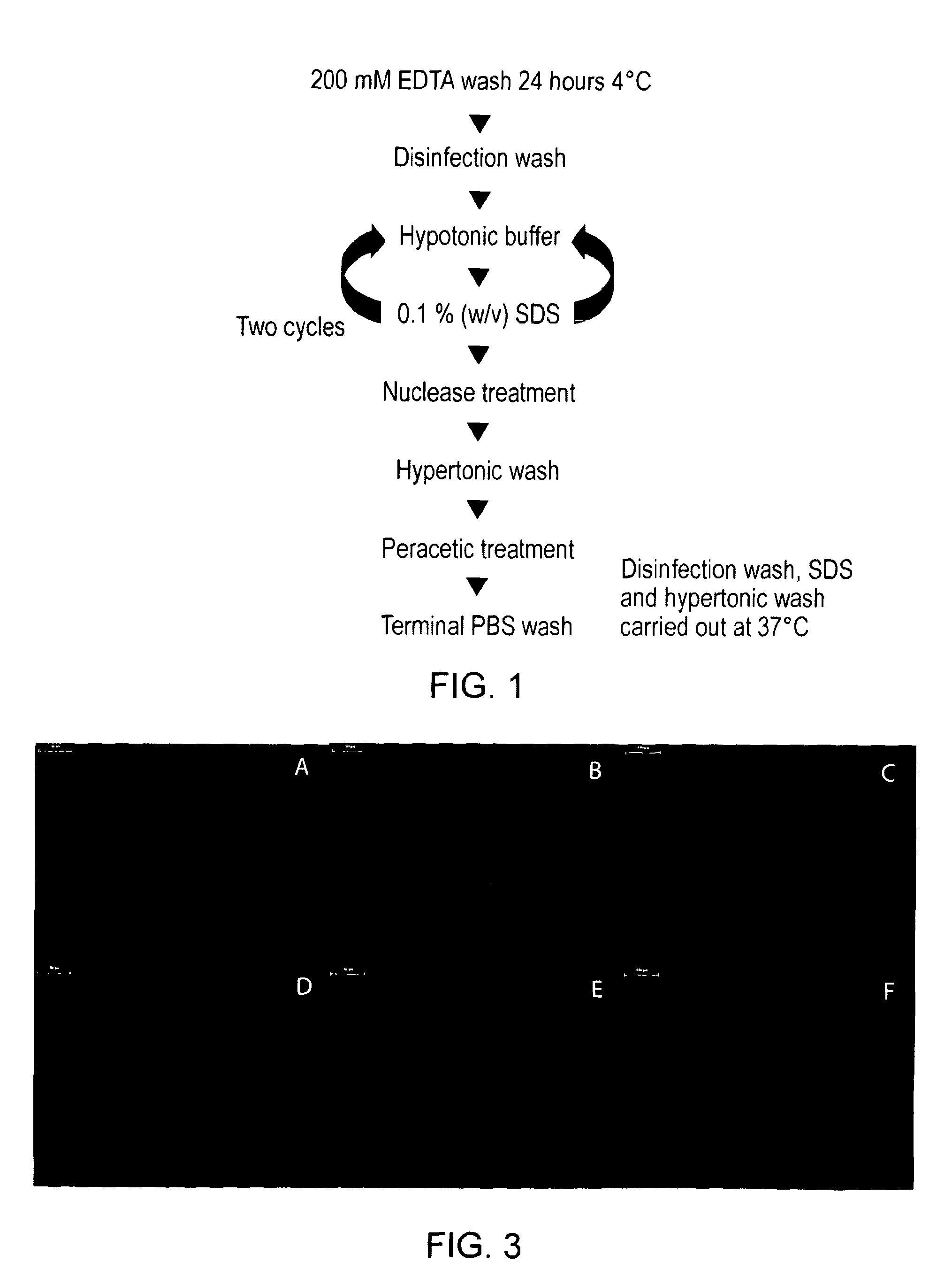
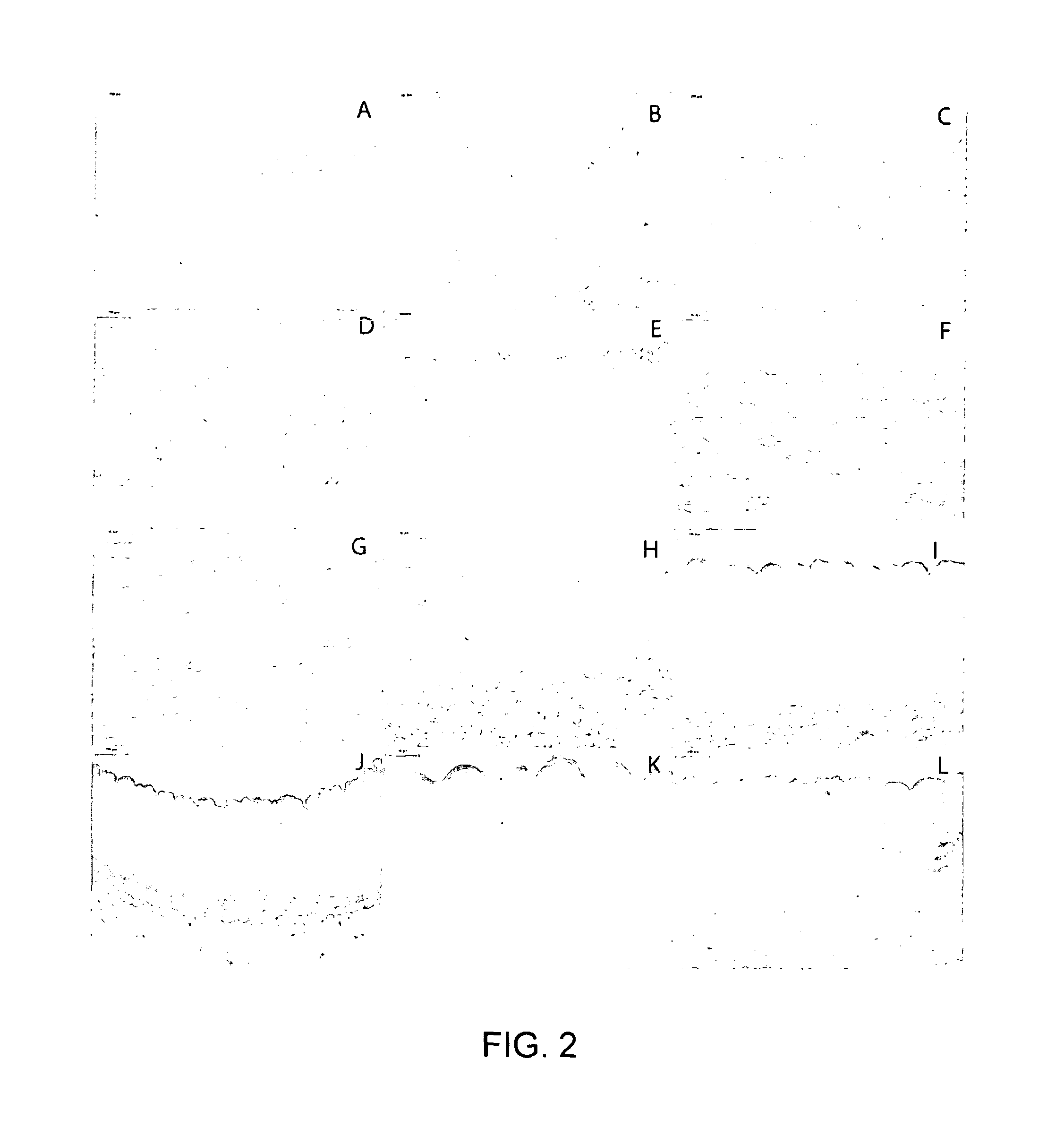
[0097]Formalin fixed treated porcine ICA and EIA was sectioned at 5 μm and stained using haematoxylin and eosin (FIG. 2). When two cycles of hypotonic buffer and SDS were used there was no evidence of residual cells or cellular remnants (FIG. 2). Additionally the matrix histoarchitecture appeared to have remained intact following treatment. Sections of formalin fixed treated porcine ICA and EIA were stained for the presence of double stranded DNA using DAPI, fresh porcine ICA and EIA were used as positive controls (FIG. 3). The staining demonstrated a lack of double stranded DNA and cells within the matrix compared to fresh control tissue (FIG. 3). When the exposure time was increased by a factor of ten no fluorescence was apparent as a result of the presence of double stranded DNA and therefore cells.
example 2
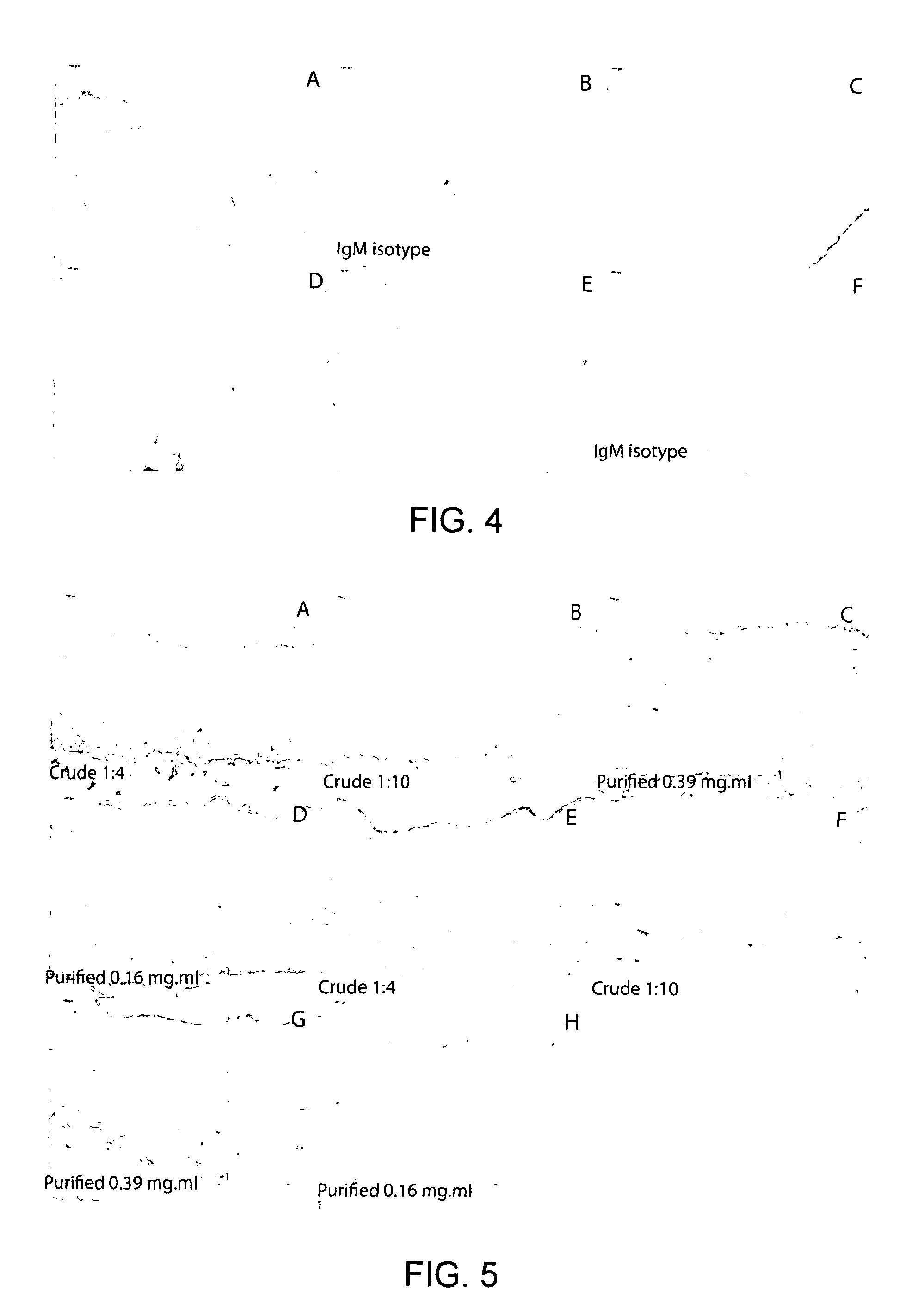
[0098]It is desirable that acellular xenogeneic matrices are devoid of the α-Gal epitope if they are to be used clinically in order to mitigate against antibody mediated inflammatory reactions. Therefore it is necessary to develop a reliable method to detect its presence within the acellular vessels. Samples of fresh and acellular porcine EIA (two cycles SDS) were formalin fixed, embedded into paraffin wax and sectioned at 5 μm. Sections of fresh and acellular porcine EIA were labelled for the presence of the α-Gal epitope using an IgM monoclonal antibody (Alexis 801-090, clone M86, dilution 1:10). The Dako Envision kit was used to visualise the primary antibody (FIG. 4). The results demonstrated the presence of the α-Gal epitope following decellularisation. There was also a high degree of background staining; this was absent when an IgM isotype control antibody was used to label the samples. Further experiments showed that the fresh tissue demonstrated good positive labelling with ...
example 3
[0099]In order to determine if there was residual α-Gal present within the acellular vessels it was important to determine the specificity of the antibody labelling. Samples of fresh and acellular EIA and ICA along with fresh and acellular α-galactosidase treated EIA and ICA were labelled for the presences of the α-Gal epitope using an IgM monoclonal antibody (Alexis ALX-801-090) clone M86 and visualised using the Dako Envision kit. Three different preparations of the antibody were used: (i) purified antibody (1:4 dilution-0.39 mg·ml−1) (ii) purified antibody adsorbed using BSA and (iii) purified antibody adsorbed using α-Gal BSA. FIG. 6 shows acellular EIA labeled using a monoclonal antibody against the α-Gal epitope, purified (A), adsorbed against BSA (B) or adsorbed against α-Gal BSA (C) or an IgM isotype control (D) with original magnification×100. FIG. 7 shows the same but for acellular ICA.
[0100]Antibody labelling of fresh tissue demonstrated strong defined positive labelling ...
PUM
| Property | Measurement | Unit |
|---|---|---|
| length | aaaaa | aaaaa |
| length | aaaaa | aaaaa |
| internal diameter | aaaaa | aaaaa |
Abstract
Description
Claims
Application Information
 - R&D
- Intellectual Property
- Life Sciences
- Materials
- Tech Scout
- Unparalleled Data Quality
- Higher Quality Content
- 60% Fewer Hallucinations
Browse by: Latest US Patents, China's latest patents, Technical Efficacy Thesaurus, Application Domain, Technology Topic, Popular Technical Reports.
© 2025 PatSnap. All rights reserved.Legal|Privacy policy|Modern Slavery Act Transparency Statement|Sitemap|About US| Contact US: help@patsnap.com


