
Method for brain tumor segmentation in multi-parametric image based on statistical information and multi-scale structure information

a brain tumor and statistical information technology, applied in the field of brain medical imaging, can solve the problems of reducing the segmentation performance, reducing the accuracy of tumor segmentation, so as to achieve accurate and reliable tumor segmentation and alleviate performance degradation

Inactive Publication Date: 2014-12-09
INST OF AUTOMATION CHINESE ACAD OF SCI
View PDF16 Cites 13 Cited by
  • Summary
  • Abstract
  • Description
  • Claims
  • Application Information

AI Technical Summary

Benefits of technology

The present invention allows for reliable and accurate tumor labeling in a compact and robust way. A label information propagation framework helps compensate for image inconsistency between training images and segments. Overall, this invention helps improve the performance and reliability of tumor segmentation.

Problems solved by technology

However, manual segmentation of brain tumor images commonly used in clinics is time-consuming, labor-intensive and subjective to considerable variation in intra- and inter-rater performance.
Supervised classification based methods with spatial regularization achieve promising performance, however, inconsistency between images to be segmented and training images will degrade the segmentation performance.
The seed selection in 3D image is time-consuming, and not easy when the boundary of target region is fuzzy.

Method used


Image

  • Method for brain tumor segmentation in multi-parametric image based on statistical information and multi-scale structure information
  • Method for brain tumor segmentation in multi-parametric image based on statistical information and multi-scale structure information
  • Method for brain tumor segmentation in multi-parametric image based on statistical information and multi-scale structure information

Examples


Embodiment Construction

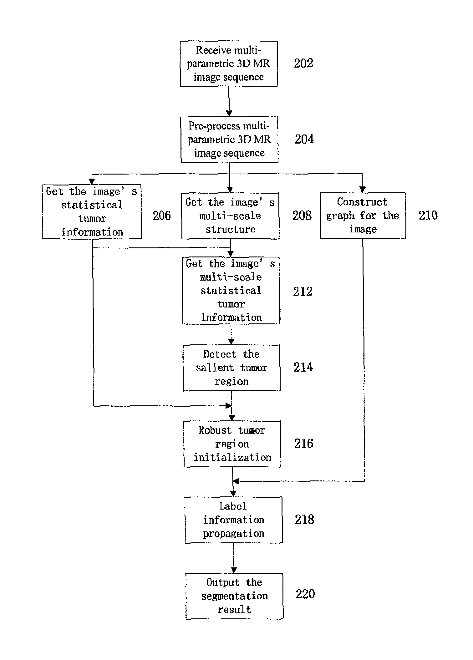
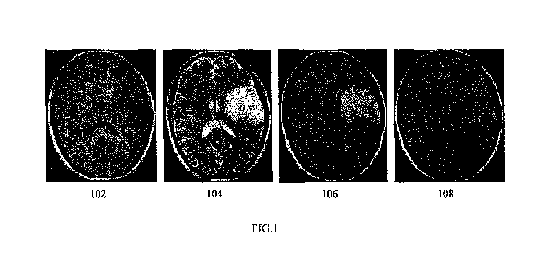
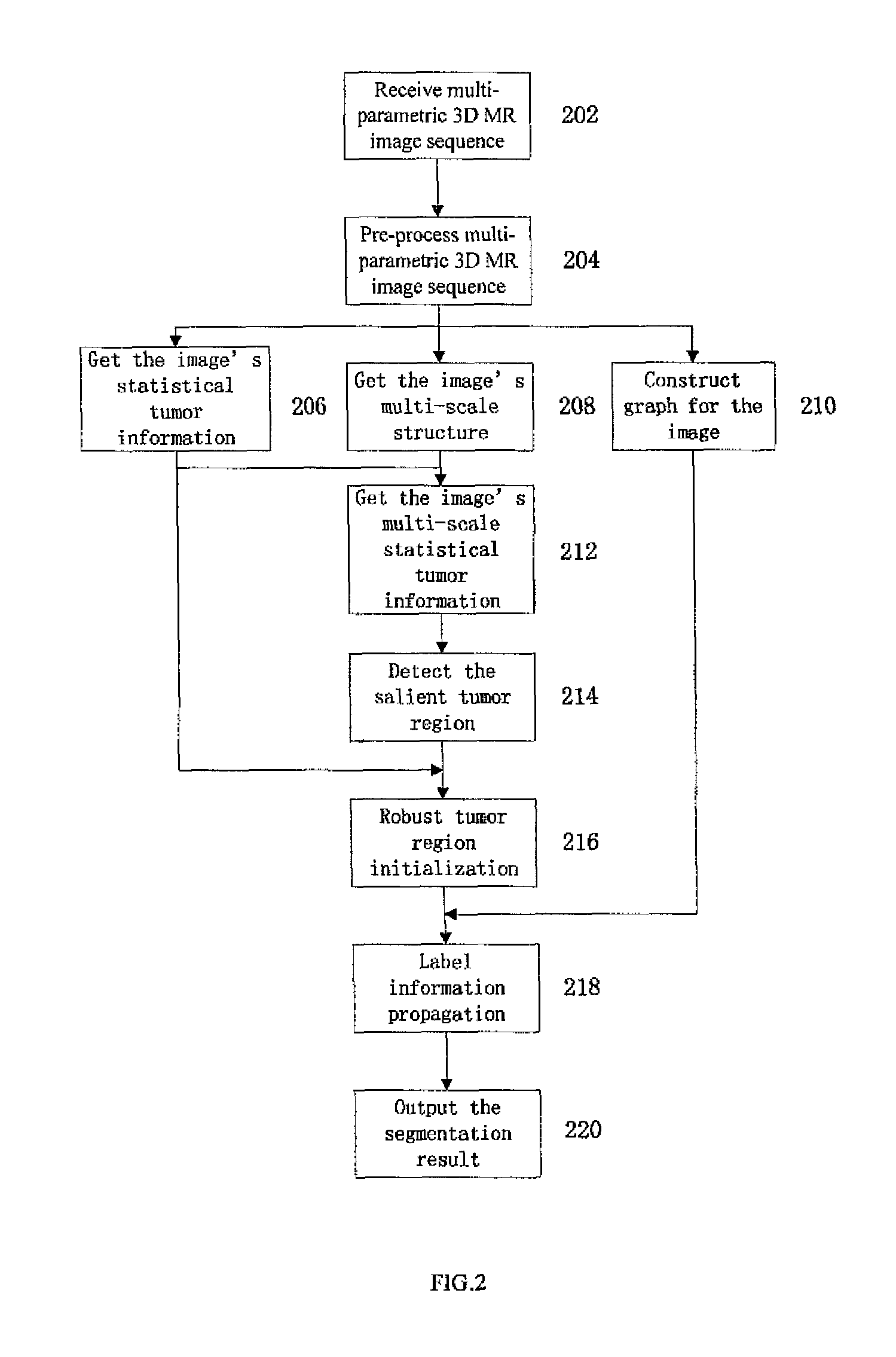
[0019]Embodiments of the present invention are directed to fully automatic brain tumor segmentation in multi-parametric 3D MR images. A multi-parametric 3D image sequence can include T1 weighted MR volume, T2 weighted MR volume, the fluid attenuation inversion recovery (FLAIR) MR volume (or the contrast enhanced T1 weighted MR volume). FIG. 1 illustrates exemplary MR images showing brain tumor. As illustration of FIG. 1, images 102, 104, 106 show an axial slice of a multi-parametric 3D MR image sequence. In particular, image 102 shows a T1 weighted image, image 104 shows a T2 weighted image, image 106 shows a FLAIR image, and image 108 shows a an annotated ground truth of brain tumor overlaid with the FLAIR image.

[0020]The present invention utilizes both the statistical tumor information from training images and the multi-scale structure information of the image to be segmented comprehensively to identify the initial tumor and non-tumor label, and subsequently applies the label info...


PUM


Abstract

A method for brain tumor segmentation in multi-parametric 3D magnetic resonance (MR) images, comprising: determining, for each voxel in the multi-parametric 3D MR image sequence, a probability that the voxel is part of brain tumor; extracting multi-scale structure information of the image; generating multi-scale tumor probability map based on initial tumor probability at voxel level and multi-scale structure information; determining salient tumor region based on multi-scale tumor probability map; obtaining robust initial tumor and non-tumor label based on tumor probability map at voxel level and salient tumor region; and generating a segmented brain tumor image using graph based label information propagation. The present invention is capable of achieving statistical reliable, spatially compact, and robust tumor label initialization, which is helpful to the accurate and reliable tumor segmentation. And the label information propagation framework could partially alleviate the performance degradation caused by image inconsistency between images to be segmented and training images.

Description

CROSS-REFERENCE TO RELATED APPLICATION[0001]This Application is a Section 371 National Stage Application of International Application No. PCT / CN2011 / 084340 filed on Dec. 21, 2011, in Chinese, the contents of which are hereby incorporated by reference in their entirety.BACKGROUND OF THE INVENTION[0002]The present invention relates to medical imaging of the brain, and particularly to fully automatic segmentation of brain tumors in multi-parametric 3D MR images.[0003]Detection and segmentation of brain tumor in magnetic resonance images is of great importance in medical diagnosis. Reliable and precise tumor segmentation provides helpful information for surgical planning and therapy accessing. However, manual segmentation of brain tumor images commonly used in clinics is time-consuming, labor-intensive and subjective to considerable variation in intra- and inter-rater performance. Accordingly, a method for fully automatic brain tumor segmentation is desirable.[0004]Several tumor segment...

Claims


Application Information

Patent Timeline
Patent Type & Authority Patents(United States)
IPC IPC(8): G06K9/00G06T7/00
CPCG06T2207/30096G06T2207/20076G06T7/0081G06T7/0087Y10S128/922G06T2207/20081G06T2207/30016G06T2207/10088G06T7/11G06T7/143
Inventor FAN, YONGLI, HONGMING
Owner INST OF AUTOMATION CHINESE ACAD OF SCI
Learn More