Diagnostic marker
a diagnostic marker and marker technology, applied in the field of diagnostic markers, can solve the problems of not providing a sufficient therapeutic outcome, affecting the so as to achieve the effect of enhancing the contrast between the cancer tissues and normal tissues and being advantageous for early detection and treatment of digestive cancer
- Summary
- Abstract
- Description
- Claims
- Application Information
AI Technical Summary
Benefits of technology
Problems solved by technology
Method used
Image
Examples
example 1
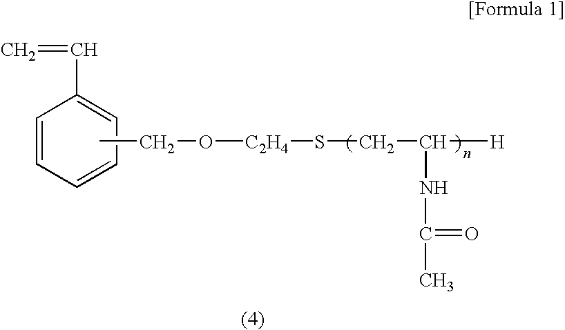
[0082]In a mixed solution of 5 mL of ion-exchanged water and 10 mL of ethanol, 1.0 g of the macro-monomer A obtained as described above, 1.0 g of styrene, and 15 mg of N,N-azobisisobutylonitrile were dissolved; after 30 minutes of nitrogen gas bubbling, the resulting solution was sealed and shaken in a 60° C. water bath for 24 hours to form fine particles.
[0083]The fine particles were centrifugally separated and freeze-dried to obtain objective fine particles. The fine particles had an average diameter of 400 nm (as measured with the dynamic light scattering method).
[0084]To 100 mg of the fine particles thus obtained, 5 mg of the fluoresceinated cholesterol obtained as described above were added. After the resulting mixture was dispersed in 5 mL of ethanol, 10 times in volume of ion-exchanged water were further added with stirring. Then, the fine particles were centrifugally separated and dispersed again in ion-exchanged water. This separation and dispersing process was repeated thr...
example 2
[0086]In a mixed solution of 5 mL of ion-exchanged water and 10 mL of ethanol, 0.75 g of the macro-monomer A obtained as described above, 0.25 g of the macro-monomer B obtained as described above, 1.0 g of styrene, and 15 mg of N,N-azobisisobutylonitrile were dissolved; after 30 minutes of nitrogen gas bubbling, the resulting solution was sealed and shaken in a 60° C. water bath for 24 hours to form fine particles. The fine particles were centrifugally separated and freeze-dried to obtain objective fine particles. The fine particles had an average diameter of 470 nm (as measured with the dynamic light scattering method).
[0087]To 100 mg of the fine particles thus obtained, similarly to Example 1, the fluoresceinated cholesterol was incorporated as an identification material and to obtain labeled fine particles.
[0088]In 800 μL of a 0.05M-KH2PO4 aqueous solution were dispersed 10 mg of the labeled fine particles obtained above. To the resulting dispersion were added 200 μL of a 0.05 M-...
example 3
[0089]In a mixed solution of 5 mL of ion-exchanged water and 10 mL of ethanol, 0.50 g of the macro-monomer A obtained as described above, 0.50 g of the macro-monomer B obtained as described above, 1.0 g of styrene, and 15 mg of N,N-azobisisobutylonitrile were dissolved; after 30 minutes of nitrogen gas bubbling, the resulting solution was sealed and shaken in a 60° C. water bath for 24 hours to form fine particles. The fine particles were centrifugally separated and freeze-dried to obtain objective fine particles. The fine particles had an average diameter of 400 nm (as measured with the dynamic light scattering method).
[0090]With respect to 100 mg of the fine particles thus obtained, similarly to Example 2, the fluoresceinated cholesterol was incorporated as an identification material to obtain labeled fine particles.
[0091]To 10 mg of the resulting labeled fine particles, similarly to Example 2, peanut lectin was bonded on the surfaces of the labeled fine particles. In this way, a ...
PUM
| Property | Measurement | Unit |
|---|---|---|
| diameter | aaaaa | aaaaa |
| diameter | aaaaa | aaaaa |
| diameter | aaaaa | aaaaa |
Abstract
Description
Claims
Application Information
 - R&D
- Intellectual Property
- Life Sciences
- Materials
- Tech Scout
- Unparalleled Data Quality
- Higher Quality Content
- 60% Fewer Hallucinations
Browse by: Latest US Patents, China's latest patents, Technical Efficacy Thesaurus, Application Domain, Technology Topic, Popular Technical Reports.
© 2025 PatSnap. All rights reserved.Legal|Privacy policy|Modern Slavery Act Transparency Statement|Sitemap|About US| Contact US: help@patsnap.com


