
Method of predicting acute appendicitis

a technology of appendicitis and appendicitis, applied in the field of acute appendicitis prediction, can solve the problems of high mortality, complicated diagnosis, and additional challenges in the accuracy of initial diagnosis

Active Publication Date: 2013-09-17
CHILDRENS MEDICAL CENT CORP
View PDF7 Cites 2 Cited by
  • Summary
  • Abstract
  • Description
  • Claims
  • Application Information

AI Technical Summary

Benefits of technology

The present invention provides devices, kits, and methods to detect the presence of acute appendicitis in a subject by detecting the presence of specific biomarkers in a urine sample obtained from the subject. The invention uses a set of appendicitis biomarkers, including leucine α-2 glycoprotein (LRG), mannan-binding lectin serine protease (MASP2), α-1-acid glycoprotein (ORM), plasminogen (PLG), mannan-binding lectin serine protease (MASP2), zinc-α-2 glycoprotein (AZGP1), apolipoprotein D (ApoD), and α-1-antichymotrypsin (SERPINA3). The invention also provides a device that can detect multiple biomarkers simultaneously. The biomarkers are detected using a protein-binding agent that specifically binds to the biomarkers. The invention can be used as a diagnostic tool for acute appendicitis.

Problems solved by technology

Untreated, mortality is high, mainly because of peritonitis and shock.
Appendicitis is among many human diseases, for which the diagnosis is complicated by the heterogeneity of its clinical presentation.
It remains the most common surgical emergency of children, with initial diagnosis accuracy additionally challenged because of non-specific but similar symptoms of many other childhood conditions.
Delays in accurate diagnosis lead to increased mortality, morbidity, and costs associated with the complications of appendicitis.
Though variable, these improvements have been modest at best, with rates of unnecessary appendectomies and ruptures of 3-30% and 30-45%, respectively.
In addition, availability of and experience with CT limit the usefulness of this approach.
Furthermore, recently its use has been re-evaluated due to concerns of cancer risk.

Method used


Image

  • Method of predicting acute appendicitis
  • Method of predicting acute appendicitis
  • Method of predicting acute appendicitis

Examples


example 1

Materials and Methods for Example 1

[0315]Sample Collection.

[0316]Urine was collected as clean catch, mid stream specimens as part of routine evaluation of 12 children and young adults (ages 1-18 years, median 11) presenting with acute abdominal pain in the Children's Hospital Boston's Emergency Department. Upon obtaining informed consent, urine was frozen at −80° C. in 12 ml aliquots in polyethylene tubes. All samples were frozen within 6 hours of collection.

[0317]Reagents.

[0318]All reagents were of highest purity available and purchased from Sigma Aldrich unless specified otherwise. HPLC-grade solvents were purchased from Burdick and Jackson.

[0319]Urine Sedimentation.

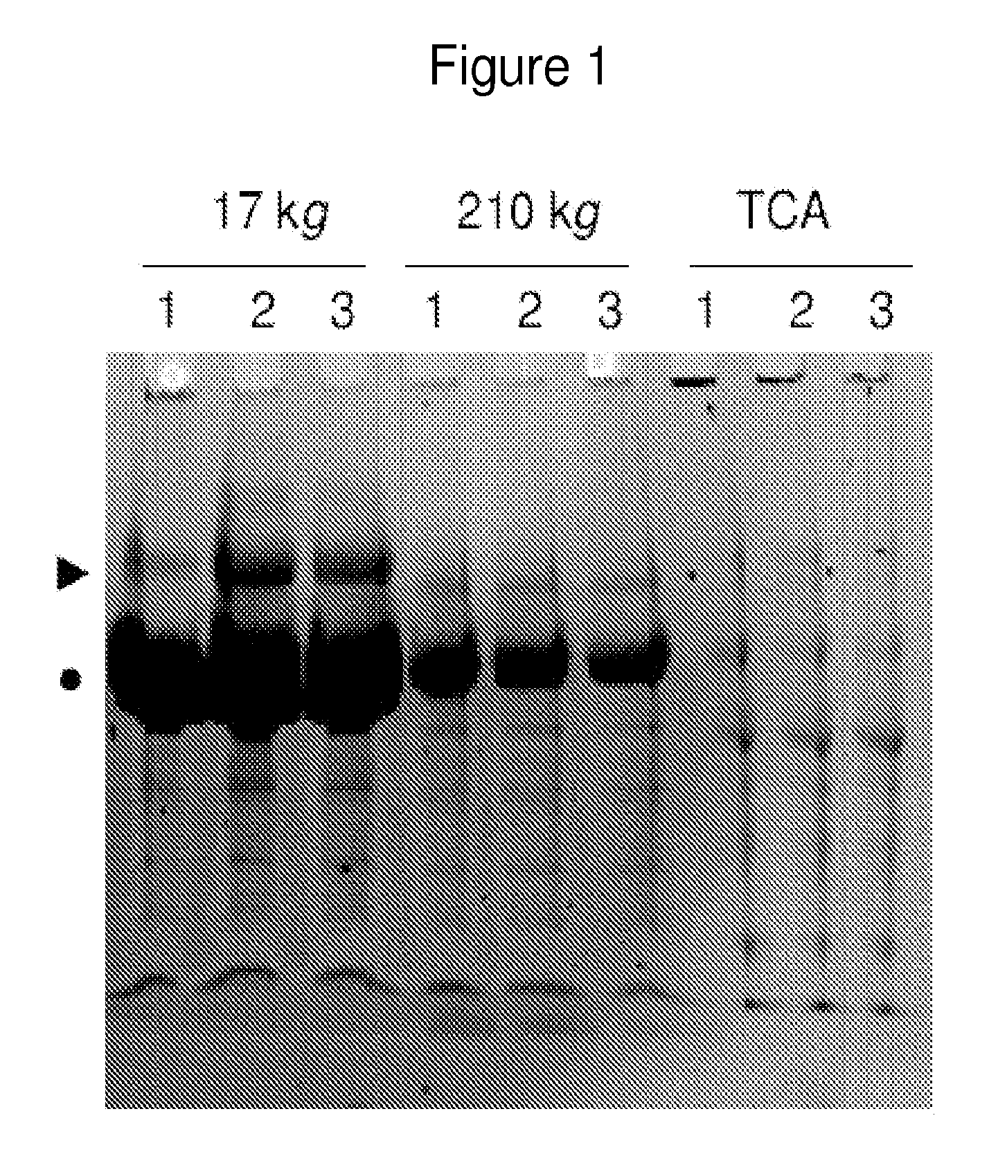
[0320]Aliquots were thawed and centrifuged at 17,000 g for 15 minutes at 10° C. to sediment cellular debris. Absence of intact cells in the sediment was confirmed by light microscopy (data not shown). Subsequently, supernatant was centrifuged at 210,000 g for 60 minutes at 4° C. to sediment vesicles and high molecular ...

example 2

Methods Use in Example 2

[0397]Study Population.

[0398]The inventors studied 67 children and young adults who presented to the ED suspected of having acute appendicitis. Patients were excluded if they had pre-existing autoimmune, neoplastic, renal or urologic disease or were pregnant. Urine was collected as clean catch, mid stream samples as part of routine ED evaluation of abdominal pain. Additional intra-individual control specimens were collected from selected patients with appendicitis after undergoing appendectomies. Informed consent was obtained prior to knowledge of final diagnosis and the urine remaining in the laboratory was retrieved and stored at −80° C. within 6 hours of collection. The expected number of patients was estimated by using the Pearson 2 test to detect a difference at a two-sided statistical significance level of 5% and power of 90% that requires 6 patients in each group, assuming that 80% of the positive samples (5 patients) will contain at least one protein ...

example 3

Discovery and Validation of Urine Markers of Acute Appendicitis Using High Accuracy Mass Spectrometry

Discovery of Diagnostic Markers by Using Urine Proteomic Profiling

[0465]In order to identify candidate urinary markers of acute appendicitis, the inventors assembled a discovery urine proteome dataset, derived from the analysis of 12 specimens, without any clinical urinalysis abnormalities, collected at the onset of the study, and distributed equally between patients with and without appendicitis. Six of these specimens were collected from patients who were found to have histologic evidence of appendicitis (2 mild, 3 moderate, 1 severe). Three specimens were collected from patients without appendicitis (1 with non-specific abdominal pain, 1 with constipation, 1 with mesenteric adenitis). From the 3 patients with appendicitis, the inventors collected additional control specimens at their routine post-surgical evaluation 6-8 weeks after undergoing appendectomies, at which time they wer...


PUM

PropertyMeasurementUnit
sizeaaaaaaaaaa
sizeaaaaaaaaaa
timeaaaaaaaaaa

Abstract

Embodiments of the invention provide method and devices for predicting the likelihood of acute appendicitis without invasive exploratory medical procedures. Several protein biomarkers: leucine-rich α-2-glycoprotein (LRG); S100-A8 (calgranulin); α-1-acid glycoprotein 1 (ORM); plasminogen (PLG); mannan-binding lectin serine protease 2 (MASP2); zinc-α-2-glycoprotein (AZGP1); Apolipoprotein D (ApoD); and α-1-antichymotrypsin (SERPINA3); are increased in the urine of patients with appendicitis. The method and devices comprise detecting the levels of these biomarkers and comparing with reference levels found in healthy individuals.

Description

CROSS REFERENCE TO RELATED APPLICATIONS[0001]This application is a 35 U. S. C. §371 National Phase Entry Application of International Application No. PCT / US2009 / 069800 filed Dec. 30, 2009, which designates the United States, and which claims benefit under 35 U. S. C. §119(e) of U. S. Provisional Application No. 61 / 141,283, filed Dec. 30, 2008, and U. S. Provisional Application No. 61 / 185,676, filed Jun. 10, 2009, the contents of which are incorporated herein by reference in their entireties.SEQUENCE LISTING[0002]The instant application contains a Sequence Listing which has been submitted in ASCII format via EFS-Web and is hereby incorporated by reference in its entirety. This ASCII copy, created on Jun. 28, 2011, is named 20110629_SequenceListing_TextFile—701039—064443_US.txt and is 244,035 bytes in size.BACKGROUND[0003]Appendicitis is a condition characterized by inflammation of the appendix. All cases require removal of the inflamed appendix, either by laparotomy or laparoscopy. U...

Claims


Application Information

Patent Timeline
Patent Type & Authority Patents(United States)
IPC IPC(8): G01N33/53G01N33/536G01N33/537G01N33/541G01N33/543
CPCG01N33/6893G01N2800/06G01N2800/50
Inventor KENTSIS, ALEXSTEEN, HANNOBACHUR, RICHARD
Owner CHILDRENS MEDICAL CENT CORP
Learn More