Cascade analysis for intestinal contraction detection
a cascade analysis and abdominal cavity technology, applied in the field of invivo imaging, can solve problems such as waste of tim
- Summary
- Abstract
- Description
- Claims
- Application Information
AI Technical Summary
Benefits of technology
Problems solved by technology
Method used
Image
Examples
example results
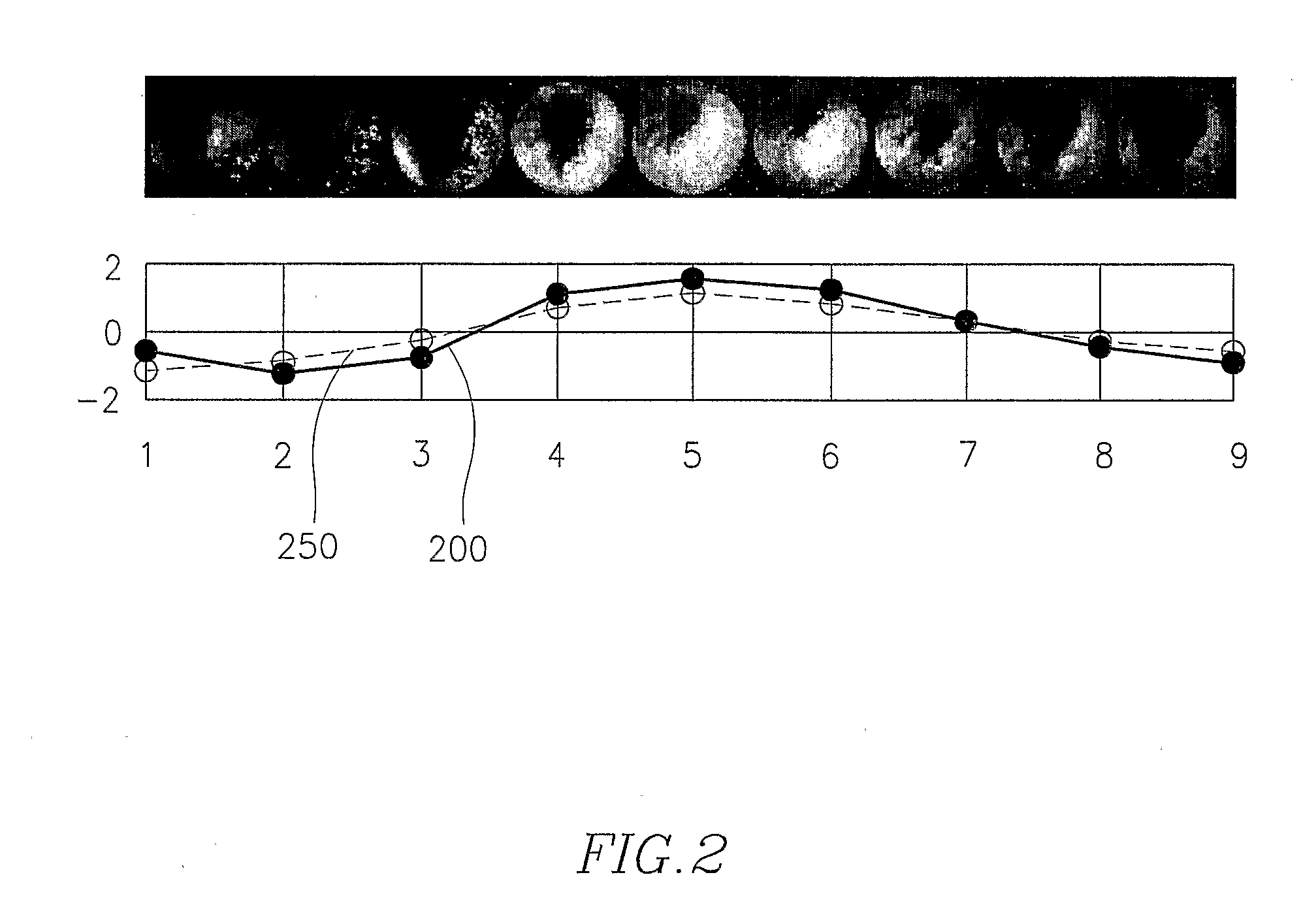
[0118]Experimental tests were performed using 10 videos obtained from 10 different fasting volunteers (without eating or drinking in the previous 12 hours to the studio), aged between 22 and 33, at the Digestive Diseases Dept. of the University Hospital Vall D'Hebron in Barcelona, Spain. The endoscope capsules used were developed by Given Imaging, Ltd., Israel. The capsules dimensions were 11×26 mm, contained 6 light emitting diodes, a lens, a colour camera chip, batteries, a radio frequency transmitter, and an antenna. The capsule acquisition rate was two frames per second. For each studio, one expert visualized the whole video and labelled all the frames showing intestinal contractions between the proximal jejunum and the first cecum images. These findings were used as the gold standard for testing the cascade system. Performance results were evaluated for each studio following the leave-one-out strategy: one video was separated for testing while the 9 remaining videos were used f...
experimental study i
[0131]Fifteen patients with chronic intestinal pseudo-obstruction (9 F, 6 M; 25-64 yrs age range) diagnosed by manometric criteria (n=9), full thickness biopsy (n=2) or both (n=4). Fifteen healthy subjects (6 F, 9 M; 19-30 yrs age range). Pillcam capsule (Given Imaging) was administered to all the participants.
[0132]The images of the small bowel emitted by the Pillcam capsule were recorded by external detectors fixed to the abdominal wall. When the capsule reached the jejunum, a test meal (Ensure HN 300 ml, 1 kcal / ml) was ingested.
[0133]Patients generally feature: 1. reduced contractility, 2. slower velocity, 3. frequent images of static tunnel reflecting quiescence, 4. more static luminal fluid and 5. impaired postprandial response with blunted postprandial increment in contractility and propulsion (*p<0.05).
[0134]Parameters were combined to achieve optimal discrimination between patients and controls.
[0135]Best discrimination was obtained when at least 2 variables were outside the...
experimental study 2
[0136]Twenty six healthy subjects were studied modelling two experimental conditions:
[0137]prokinetic stimulation, induced by ingestion of a meal (Ensure HN, 300 ml, 1 kcal / ml; n=13)
[0138]motor inhibition, induced by glucagon administration (4.8 μg / kg bolus plus 9.6 μg / kg h infusion for 1 h; n=5)
[0139]basal conditions, as control (fasting; n=8)
[0140]The meal stimulus increased the number of contractions, while motor inhibition suppressed contractile activity as compared to basal conditions. Propulsion of the capsule increased by 118% during prokinetic stimulation, and decreased by 73% during inhibition. During motor inhibition the percentage of time when the capsule was completely still was much longer than during basal conditions. Conversely, prokinetic stimulation reduced the time during which the gut presented a static tunnel appearance. Prokinetic stimulation reduced the tone of appearance of endoluminal secretions.
PUM
 Abstract
Description
Claims
Application Information
 - R&D
- Intellectual Property
- Life Sciences
- Materials
- Tech Scout
- Unparalleled Data Quality
- Higher Quality Content
- 60% Fewer Hallucinations
Browse by: Latest US Patents, China's latest patents, Technical Efficacy Thesaurus, Application Domain, Technology Topic, Popular Technical Reports.
© 2025 PatSnap. All rights reserved.Legal|Privacy policy|Modern Slavery Act Transparency Statement|Sitemap|About US| Contact US: help@patsnap.com


