Porous material having hierarchical porous structure and preparation method thereof
a porous structure and porous material technology, applied in the field of porous ceramic balls with hierarchical porous structure, can solve the problems of difficult control of pore size, poor interporous open structure of porous materials obtained by conventional methods, and pore plugging, etc., to achieve the effect of increasing viscosity
- Summary
- Abstract
- Description
- Claims
- Application Information
AI Technical Summary
Benefits of technology
Problems solved by technology
Method used
Image
Examples
example 1
Porous Ceramic Balls 1
[0068]Step 1. Preparation of Polymer Template Solution
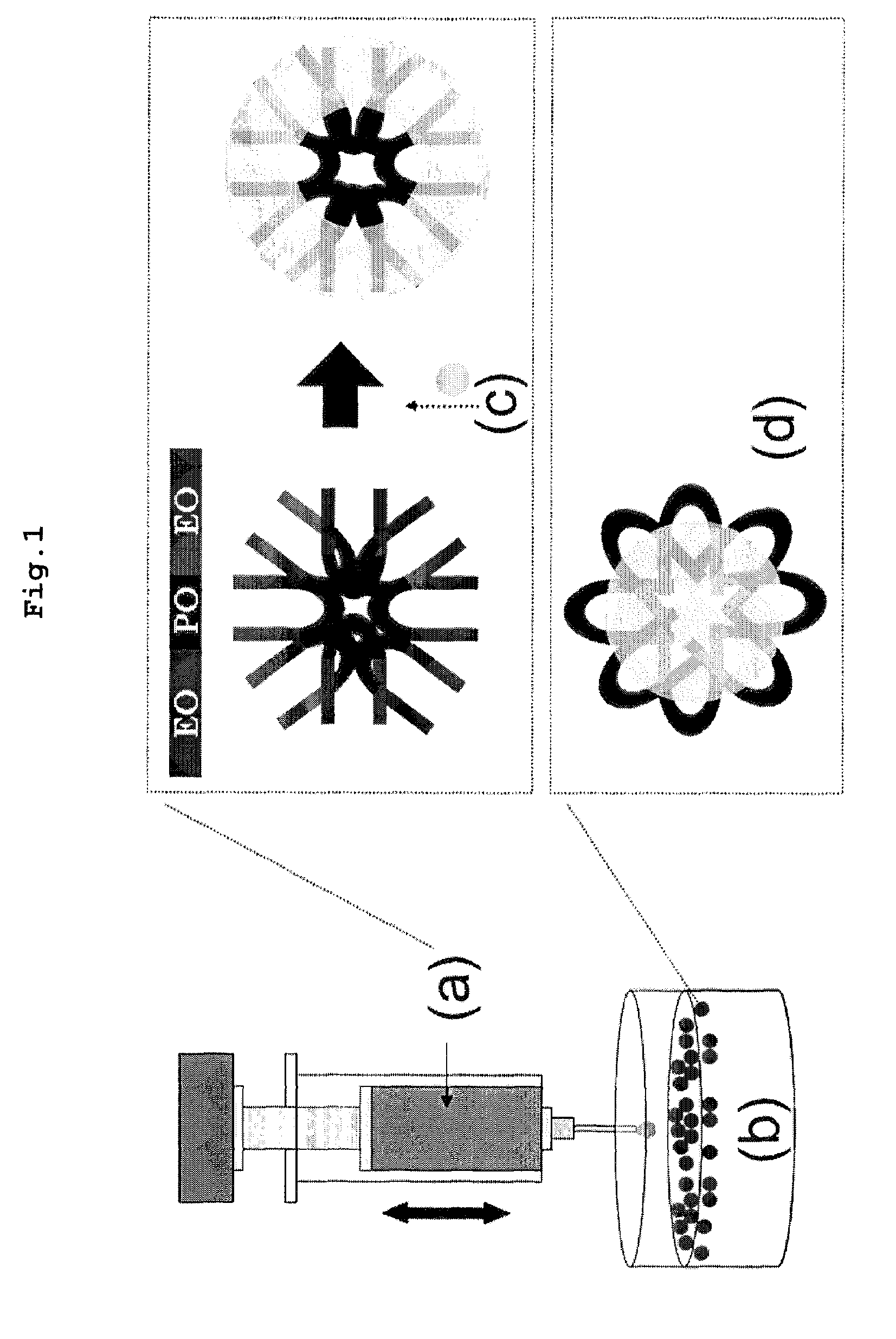
[0069]3.46 g of the tri-block copolymer (Poly-ethylene oxide)132 (Poly-propylene oxide)50 (Poly-ethylene oxide)132 was completely dissolved in 18.1 mL of ethanol at 40° C. for 0.5 to 1 hour with stirring, to afford a polymer template solution.
[0070]Step 2. Preparation of Precursor Solution
[0071]6 mL of tetraethyl orthosilicate was slowly mixed with 1.36 g of calcium nitrate tetrahydrate to homogeneity, followed by the addition of 0.26 mL of triethyl phosphate. Then, a mixture of 0.95 mL of 1 M HCl, 7.62 mL of ethanol and 2.86 mL of distilled water was added before stirring at 40° C. for 0.5-1 hour. Thus, a precursor solution resulted.
[0072]Step 3. Preparation of Mix Solution for Porous Ceramic Balls
[0073]To the solution of step 1 was added a B solution with slowly stirring. The resulting mixture was further stirred at 1000-1500 rpm for 2-4 hours and then incubated at −5 to 80° C. at RH 5˜1000 for 24˜48 hours...
example 2
Porous Ceramic Balls 2
[0080]The same procedure as in Example 1 was repeated, with the exception that (Poly-ethylene oxide)100(Poly-propylene oxide)65(Poly-ethylene oxide)100 was used instead of the tri-block copolymer of step 1 of Example 1.
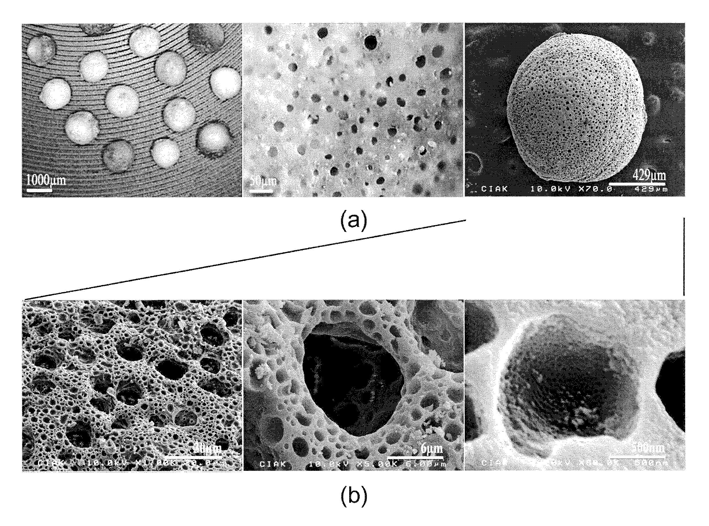
[0081]Referring to FIG. 4, scanning electron microscope photographs of the porous ceramic balls are shown.
[0082]As seen in these SEM photographs, the porous ceramic balls of Example 2 were found to be similar in morphology to those of Example 2.
example 3
Preparation of Bioactive Porous Ceramic Balls
[0083]The porous ceramic balls of Example 1 were immersed for 1, 4 and 24 hours in simulated body fluid to allow apatite crystals to grow therein. The SEM photographs of the porous ceramic balls with apatite crystals grown therein are given in FIG. 12.
[0084]As seen in these SEM photographs, apatite crystals are formed even after immersion for 1 hour in simulated body fluid, in comparison to no apatite crystals before immersion in the simulated body. Further, following lapses of 4 and 24 hours, apatite crystals in the simulated body fluid uniformly grow over the surfaces of the hierachically interconnected pores within the porous ceramic balls. In addition, the apatite crystals are in tens of nanometers of size, similar to the dimension of actual bones, with a needle-like morphology.
PUM
| Property | Measurement | Unit |
|---|---|---|
| size | aaaaa | aaaaa |
| size | aaaaa | aaaaa |
| size | aaaaa | aaaaa |
Abstract
Description
Claims
Application Information
 - R&D
- Intellectual Property
- Life Sciences
- Materials
- Tech Scout
- Unparalleled Data Quality
- Higher Quality Content
- 60% Fewer Hallucinations
Browse by: Latest US Patents, China's latest patents, Technical Efficacy Thesaurus, Application Domain, Technology Topic, Popular Technical Reports.
© 2025 PatSnap. All rights reserved.Legal|Privacy policy|Modern Slavery Act Transparency Statement|Sitemap|About US| Contact US: help@patsnap.com


