
Molecules for targeting compounds to various selected organs or tissues

a technology of compounds and molecules, applied in the field of in vivo targeting, can solve the problems of poor in vivo muscle uptake of these aons, unsatisfactory side effects, and the biggest hurdle to overcome, and achieve the effect of improving in vivo uptak

Active Publication Date: 2012-09-18
BIOMARIN TECH BV
View PDF134 Cites 20 Cited by
  • Summary
  • Abstract
  • Description
  • Claims
  • Application Information

AI Technical Summary

Benefits of technology

The use of muscle-specific peptides and peptidomimetics significantly improves the uptake of therapeutic agents into muscle cells, including the heart, leading to enhanced treatment efficacy for muscle diseases and reduced systemic toxicity, as demonstrated by increased dystrophin expression and exon skipping in muscle tissues.

Problems solved by technology

This can result in undesirable side effects due to, for example, generalized toxic effects throughout the patient's body.
However, the biggest hurdle to overcome is the poor in vivo muscle uptake of these AONs, especially in all kind of myopathies like Myotonic Dystrophy (MD) and Spinal Muscular Atrophy (SMA).
None of the mechanisms investigated to date have the ability to specifically deliver (antisense) oligonucleotides, let alone entire genes, to essentially all muscle tissues / cells simultaneously over the entire body.
However, even after repeated administration, dystrophin expression was barely detectable in diaphragm and was undetectable in heart muscle.
Furthermore, in these mdx mice the AONs are taken up rather easy into the muscles because the muscle membranes are compromised, which is not the case for the muscles of, for instance, young Duchenne patients.
Also, in other muscle diseases like SMA and MD delivery of AON is complicated due to the fact that in this case the muscle cell walls are not compromised.
However, it has not yet been shown whether or not this peptide can be used for in vivo targeting of conjugated compounds to muscle cells.
Also here it has not yet been shown whether or not these peptides can be used for in vivo targeting of conjugated compounds to muscle cells.
In the case of lysosomal storage disease a problem in the enzyme replacement therapy is poor in vivo uptake of the therapeutic recombinant enzyme into the muscle cells.

Method used


Image

  • Molecules for targeting compounds to various selected organs or tissues

Examples


example 1

In vitro Selection of Peptides Against Myoblasts and Myotubes

[0081]A pre-made phage peptide library containing 2 billion phages expressing random 7-mer peptides (New England Biolabs Inc.) has been screened to identify muscle-specific peptides. Briefly, the library of phage-displayed peptides was incubated with cells plated in culture flasks. After washing away the unbound phage, specifically-bound or internalized phage was eluted and amplified. After a series of different in vitro biopanning steps, including both positive and negative selection rounds on human or mouse myotubes and fibroblasts respectively, the pool was enriched for binding sequences which could be characterized by DNA sequencing. Therefore, muscle-specific peptides were identified which will bind to and be internalized by the target cells. Specific peptide sequences that were found are shown in Table 1.

[0082]

TABLE 1Peptide sequences found after in vitro selection on human and mouse myotubesSPNSIGT1STFTHPR1QLFTSAS1S...

example 2

Selection of Peptides in mdx Mice

[0085]For panning experiments in mdx mice, the library was injected through the tail vein. After 10 to 20 minutes, the mice were sacrificed and perfused, after which the heart and different muscle groups were isolated. Bound and / or internalized phage was recovered from tissue homogenates, amplified, and re-applied to mdx mice. Enriched sequences were selected and further characterized. Specific peptide sequences that were found are shown in Table 2.

[0086]

TABLE 2Peptide sequences found after four rounds of in vivo selection in mdx mice, on skeletal muscle and heartYQDSAKT1,2VTYKTASEPLQLKM1WSLQASHTLWVPSR1(SEQ ID NO.: 7)(SEQ ID NO.: 80)(SEQ ID NO.: 81)(SEQ ID NO.: 82)ISEQ ID NO.: 83)QGMHRGTLYQDYSL1SESMSIKLPWKPLG1QSPHTAP(SEQ ID NO.: 84)(SEQ ID NO.: 85)(SEQ ID NO.: 86)(SEQ ID NO.: 87)(SEQ ID NO.: 88)TPAHPNY1SLLGSTPTALPPSY1VNSATHSLPLTPLP1(SEQ ID NO.: 89)(SEQ ID NO.: 90)(SEQ ID NO.: 91)(SEQ ID NO.: 92)(SEQ ID NO.: 93)NQLPLHAGNTPSRA1,2TQTPLKQAMISAIHNLTRLHT(S...

example 3

In vivo Staining of Muscle Fibers After Intramuscular Injection

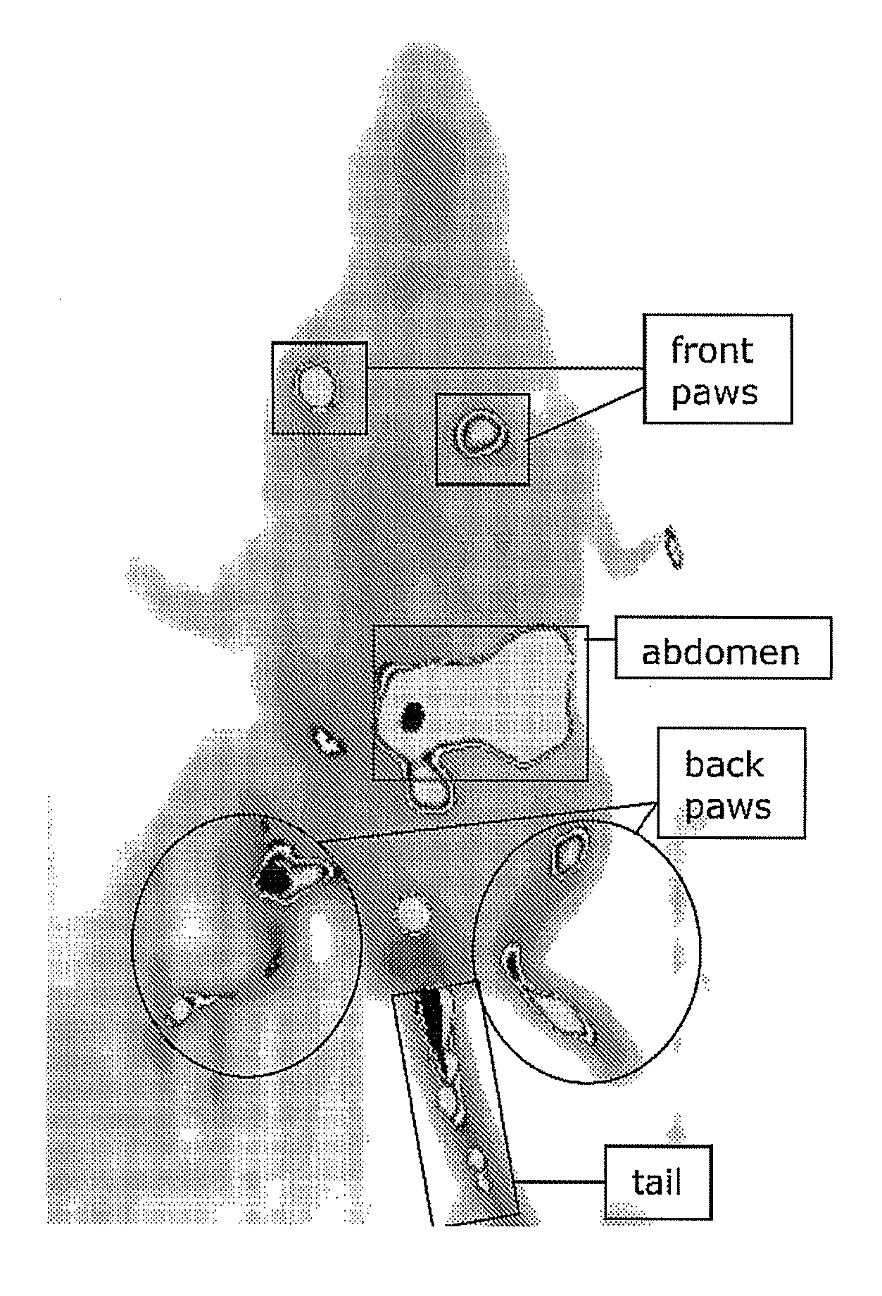
[0088]Peptides that showed uptake into cultured human myoblasts and myotubes were synthesized with a fluorescent label (FAM) and injected into the gastrocnemius (calf muscle) of four week old mdx mice. FAM-labeled peptides QLFTSAS (SEQ ID NO.: 3) (5 nmol injected), LYQDYSL (SEQ ID NO.: 85) (2.5 nmol injected) and LGAQSNF (SEQ ID NO.: 100) (2.5 nmol injected) were analysed. After three days the mice were sacrificed and the muscles were snap frozen. Cross-sections were cut, fixed with acetone and mounted for analysis with a fluorescence microscope. Cross-sections were cut and photographed with a fluorescence microscope (CCD camera).

[0089]The photographs showed that the peptides QLFTSAS (SEQ ID NO.: 3), LYQDYSL (SEQ ID NO.: 85) and LGAQSNF (SEQ ID NO.: 100) were taken up into a large area of muscle fibers and were still visible after 3 days. It was clearly shown that whole fibers are stained homogeneously, and that sometime...


PUM

PropertyMeasurementUnit
pharmaceutical compositionaaaaaaaaaa
chiralityaaaaaaaaaa
lengthaaaaaaaaaa

Abstract

The invention provides conjugates, comprising an organ, tissue or tumor cell homing molecule linked to a moiety. Such a moiety can be, for example, an oligonucleotide, small interfering RNA, gene, virus, protein, pharmaceutical or detectable agent. In addition the invention provides methods to diagnose or treat a pathology of the muscle or heart, by administrating to a subject having or suspected of having a pathology a molecule or conjugate that homes to, binds to and is taken up by the muscle cells or heart cells.

Description

RELATED APPLICATIONS[0001]This present invention is a continuation patent application that claims priority to PCT patent application number PCT / NL2008 / 050475, filed Jul. 14, 2008, and European patent application No. 07112323.7, filed on Jul. 12, 1007, the entirety of which are herein incorporated by reference.FIELD OF THE INVENTION[0002]The present invention is in the field of in vivo targeting and provides molecules that home to, bind to and are taken up by various organs or tissues.BACKGROUND OF THE INVENTION[0003]Most therapeutic compounds are delivered to the target organ or tissue through the circulation. However, in most cases the drug or other treatment will not only target the diseased organ or tissues, but will also be taken up by other organs and tissues in the body. This can result in undesirable side effects due to, for example, generalized toxic effects throughout the patient's body. Thus, it would be desirable to selectively target specific organs or tissues. In additi...

Claims


Application Information

Patent Timeline
Patent Type & Authority Patents(United States)
IPC IPC(8): C07K17/02
CPCA61K47/48238A61K49/0056A61K49/0047A61K49/0032A61K47/62A61P19/00A61P21/00A61P21/02A61P21/04A61P3/00A61P3/10A61P3/04A61P5/00A61P9/00
Inventor HEEMSKERK, JOHANNES ANTONIUSVAN DEUTEKOM, JUDITH CHRISTINA THEODORAVAN KUIK-ROMEIJN, PETRAPLATENBURG, GERARD JOHANNES
Owner BIOMARIN TECH BV
Learn More