Method and apparatus for connective tissue treatment

a connective tissue and treatment method technology, applied in the direction of elcosanoid active ingredients, peptide/protein ingredients, therapy, etc., can solve the problems of reducing the healing time, reducing the applicability of utility, and not having comparable proteoglycan content, etc., to achieve the effect of promoting healing and wide applicability

Inactive Publication Date: 2012-02-28
EXOGEN
View PDF400 Cites 118 Cited by
  • Summary
  • Abstract
  • Description
  • Claims
  • Application Information

AI Technical Summary

Benefits of technology

[0024]Because of the broad applicability of utility of the invention in promoting healing, the method and apparatus described herein are useful in treating patients with a broad range of problems, such as trauma, tissue insufficiency, pain, post-surgical healing, degenerative conditions such as osteoarthritis, and other problems. Moreover, because the invention is portable, does not require prolonged treatment times, and is designed for ease of use and positioning of the ultrasonic transducers, patients will be more likely to use the technique properly and sufficiently to benefit therefrom.

Problems solved by technology

However, the resulting nonhyaline or fibrocartilage produced is biomechanically inferior to articular cartilage, does not have comparable proteoglycan content, and may consist primarily of a thin unorganized layer of collagen.
Further, it has been observed that degeneration of the new tissue generally occurs over time, requiring the need for additional reconstructive surgical treatment.
Further, there is a need for an apparatus which stimulates cartilage regeneration and significantly reduces the healing time.
However, while these approaches may control symptoms, they do not effectively address the underlying damage to connective tissue, such as cartilage.
Moreover, the drugs used may cause severe side effects in some patients, which can result in hospitalization and, in some cases, death.
However, injuries and pathologies of cartilage are not the only conditions of connective tissue requiring treatment that involve significant healing time.
When ligament and tendons rupture the patients have pain and laxity of the joint or muscle.
Increased vascularization will lead to improved, faster healing, while insufficient vascularization can lead to necrosis of the tissue.
This is a lengthy process during which time the graft loses strength and is at risk of rerupture or damage.
This leads to lengthy rehabilitation times (e.g., a minimum of 6 months for anterior cruciate ligament (ACL) reconstruction).
The phenomenon of “bone tunnel widening” can often present a problem.
However, physiotherapy is inconvenient, time consuming and relatively expensive, which can lead to problems with patient compliance.
Applying thermal energy to collagen can cause an alteration of the molecular configuration resulting in shrinkage.
This results in a shorter structure and a ‘tighter’ joint.
However, the thermal energy incidentally may also damage the tissue resulting in loss of viability of cells, loss of blood supply and reduced mechanical integrity of the structure.
Tissue shrinkage procedures have thus had difficulties resulting from decreased biomechanical integrity and long rehabilitation times due to slow healing.
This has resulted in the ‘modified’ tissue being stretched back out prior to its healing and reestablishment of normal biomechanical strength, resulting in the loss of the benefit of the shrinkage procedure.
This damage represents a significant disadvantage and has inhibited the use of capsulorraphy and related tissue shrinkage procedures.
One of the shortcomings of this approach is that it is not possible to grow a tissue-engineered material (implant, graft or organ) with a vascular supply in vitro.

Method used


Image

  • Method and apparatus for connective tissue treatment
  • Method and apparatus for connective tissue treatment
  • Method and apparatus for connective tissue treatment

Examples


example 1

[0115]An ovine model of soft tissue graft (digital extensor tendon) reconstruction of the ACL was utilized to allow for assessment of the method and apparatus of the invention. A modified ACL reconstruction was performed on 21 animals in three groups of 7 animals. Control groups of 2 animals (provided with no ultrasound treatment), and experimental groups of animals (provided with ultrasound treatment for 20 minutes continuously each day) were harvested at the end of 3, 6, and 12 weeks.

[0116]The right hind limb was operated on in all animals. The anterior cruciate ligament (ACL) was visualized and removed at the insertion site through an antero-medial arthrotomy. The ACL reconstruction was performed using a digital extensor tendon graft. The tendon graft was harvested from the same limb by 2 stab incisions. The graft was whip stitched using # 2 Ethibond and prepared using the Acufex Graftmaster. The graft was doubled and passed through 4.5 mm tunnels in the tibia and femur. Endobutt...

example 2

[0122]Hartley strain guinea pigs that spontaneously develop osteoarthritis (OA) were used for this study. This strain develops an arthropathology that mimics human OA between the age of 6 and 12 months of age. The OA is confined to the medial tibia plateau in the early stages of the disease.

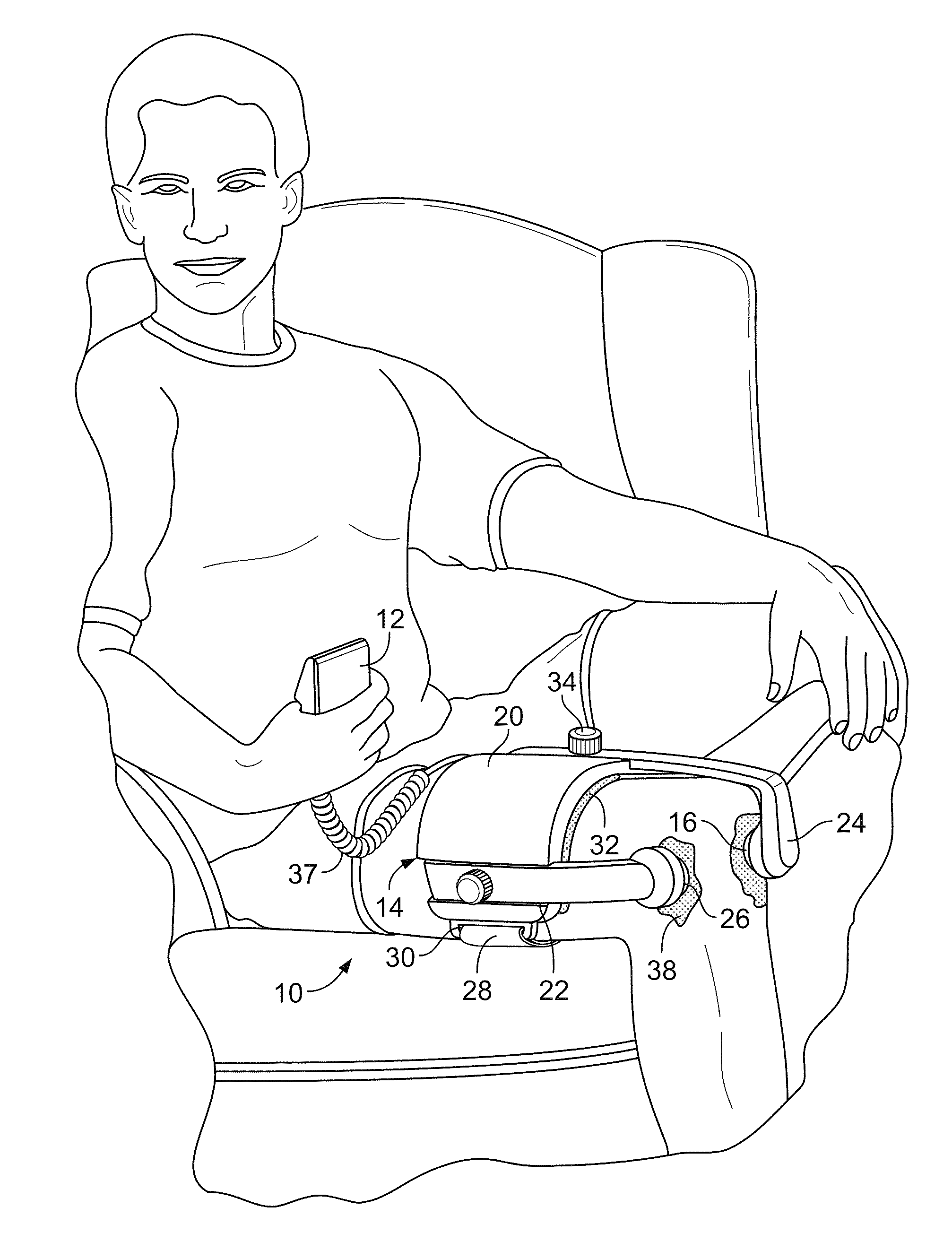
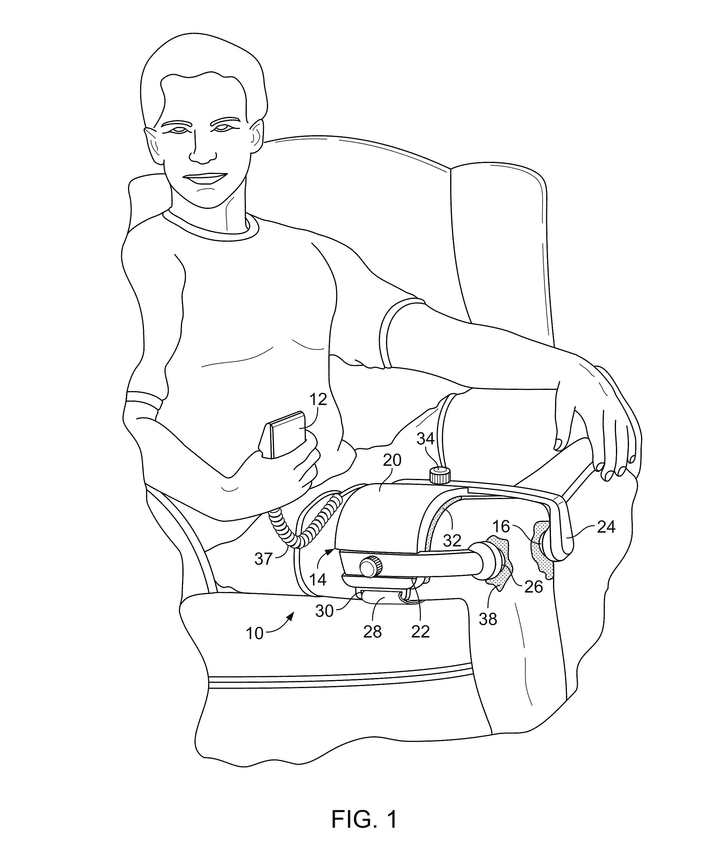
[0123]Eight animals were utilized for this study. The animals were 2 months of age when the study was initiated. The left legs of the animals were treated with pulsed low intensity ultrasound (1.5 MHz, pulsed at 1 KHz, 200 μs burst width) for a period of 4 months. The ultrasound was applied for 20 minutes per day, for 5 days per week. The ultrasound transducer was coupled to the skin with gel on the media side of the left knee after first shaving the knee joint. The transducers were held in place during treatment with strapping. The animals were terminated at 6 months of age after 4 months of treatment.

[0124]The ultrasound treated (FIG. 23A) and control (FIG. 23B) knee joints were dissected and d...


PUM


Abstract

The invention relates to methods and apparatus for therapeutically treating connective tissue or increasing vascularization in tissue using ultrasound. More particularly, the present invention relates to methods and apparatus which use ultrasound to stimulate growth or healing, or to treating pathologies, of connective tissue, or to increase vascularization in ischaemic or grafted tissue using ultrasound.

Description

CROSS-REFERENCE TO RELATED APPLICATIONS[0001]This application is a continuation application, of and claims priority to, U.S. application Ser. No. 10 / 131,784, filed Apr. 24, 2002, now pending, which is a continuation of U.S. Ser. No. 10 / 096,216, filed Mar. 11, 2002, now abandoned, which is a continuation of Ser. No. 09 / 436,999, filed Nov. 9, 1999, now U.S. Pat. No. 6,355,006, which is a continuation of International Application No. PCT / US98 / 02447, filed Feb. 6, 1998, which claims priority to U.S. Provisional Application No. 60 / 037,367 filed on Feb. 6, 1997, and which is a continuation-in-part of U.S. application Ser. No. 09 / 568,481 filed May 9, 2000, now U.S. Pat. No. 6,432,070, which claims priority to U.S. Provisional Application No. 60 / 133,442, filed May 11, 1999, the entire contents of each of which are incorporated herein by reference.BACKGROUND OF THE INVENTION[0002]1. Field of the Invention[0003]The present invention relates to methods and apparatus for therapeutically treatin...

Claims


Application Information

Patent Timeline
Patent Type & Authority Patents(United States)
IPC IPC(8): A61H1/00A61H23/02A61B17/16A61B18/20A61F5/01A61F5/02A61K31/557A61K38/22A61N7/00A61P19/04A61P43/00
CPCA61N7/00A61B17/16A61B18/20A61N2007/0078A61P19/04A61P43/00
Inventor HUCKLE, JAMES WILLIAMCARTER, ANDREWCOTTON, NICOLAS J.WALSH, WILLIAM RSCARBOROUGH, NELSONTALISH, ROGER J.
Owner EXOGEN
Why Eureka
Try Eureka