
Method for regenerating cartilage

a cartilage composition and regenerative technology, applied in the field of methods, can solve the problems of enlargement and progression of lesion, limited repair potential of articular cartilage of higher animals, and enlargement of lesion, and achieve the effect of effective cartilage composition and reliable inducement of new cartilage formation

Active Publication Date: 2010-11-23
VIVEX BIOLOGICS GRP INC
View PDF21 Cites 23 Cited by
  • Summary
  • Abstract
  • Description
  • Claims
  • Application Information

AI Technical Summary

Benefits of technology

This approach induces complete regeneration of articular cartilage defects in non-human primates, showing rapid new cartilage formation and full coverage within sixteen weeks, even in defects involving subchondral bone, without altering growth factors or requiring complex extraction processes.

Problems solved by technology

Articular cartilage of higher animals, including man, has very limited potential for repair.
Following injuries to articular cartilage, the cartilage generally does not repair itself because of its very limited capacity for regeneration.
In injuries involving subchondral bone, there is no regeneration of the cartilage, but there is typically enlargement and progression of the lesion with associated pain.
The ultimate goal of treatment of these lesions is restoration of the cartilage, but in most cases this is not achieved.
However, none of these produces regeneration of the native articular cartilage.
However, it is not a cartilage repair procedure, but rather merely removes frayed cartilage components as a palliative approach that may delay or defer unicondylar or total knee arthroplasty.
However, this does not result in regeneration of articular hyaline cartilage.
This technique is limited by the size of the defect and by the amount of healthy cartilage that can be harvested from a given patient.
A drawback of the technique is the necessity to anatomically match the curvature and size of the graft with the defect in the recipient.
The technique does not produce regeneration of the native cartilage, but depends on obliterations of the defect either with autologous or allogeneic osteochondral grafts.
However, to date, application of various BMP's to chondral defects has failed to produce full-thickness repair and regeneration.
Most other studies of articular cartilage regeneration have been performed on rodents, and none of these has produced complete articular cartilage regeneration.

Method used


Image

  • Method for regenerating cartilage
  • Method for regenerating cartilage
  • Method for regenerating cartilage

Examples


example

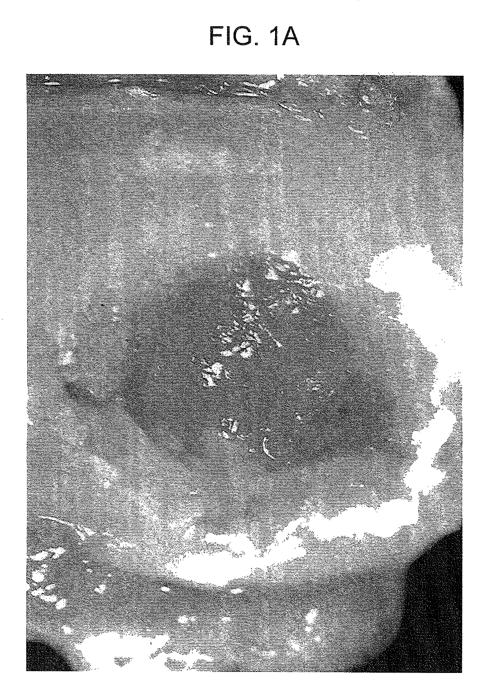
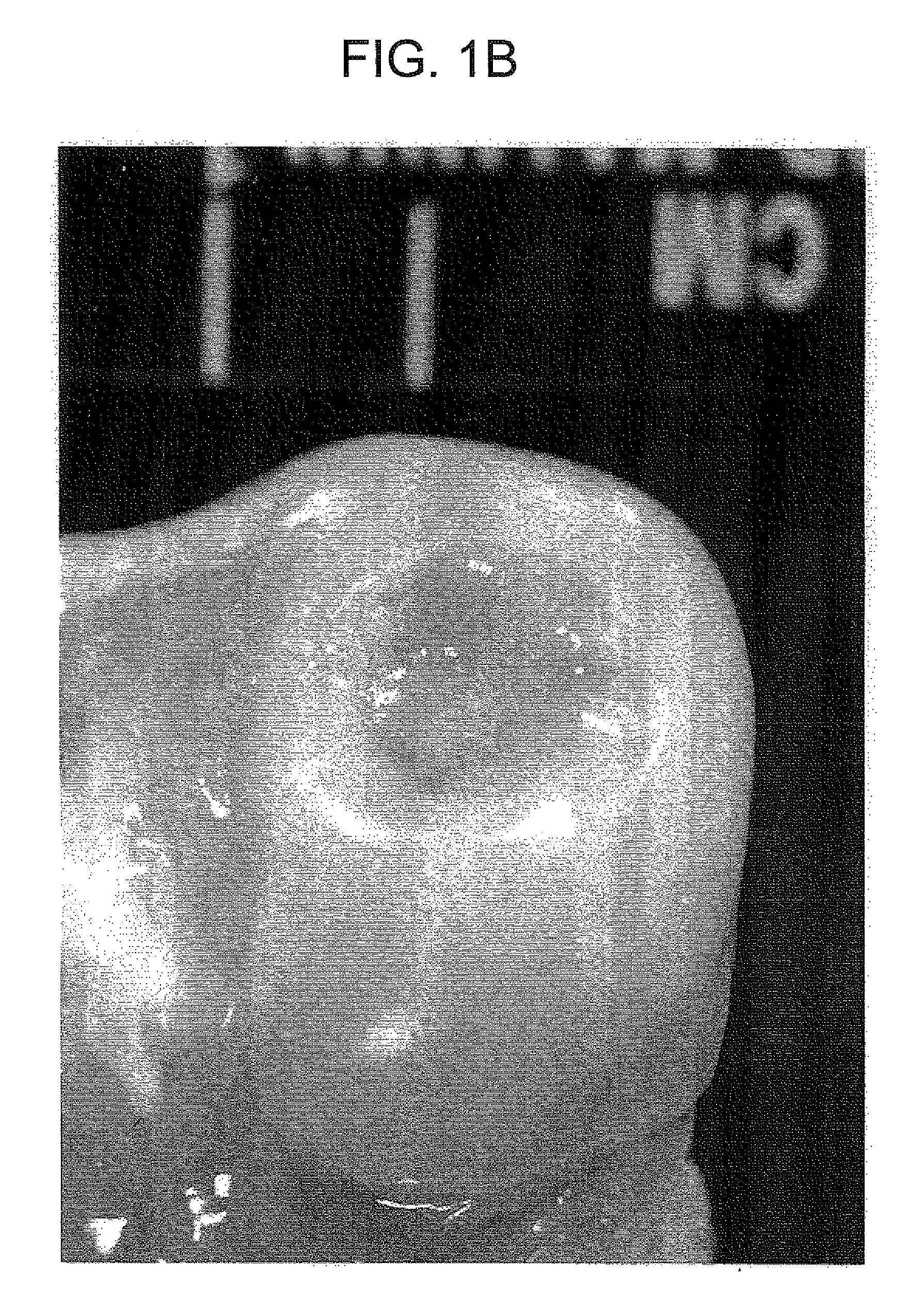
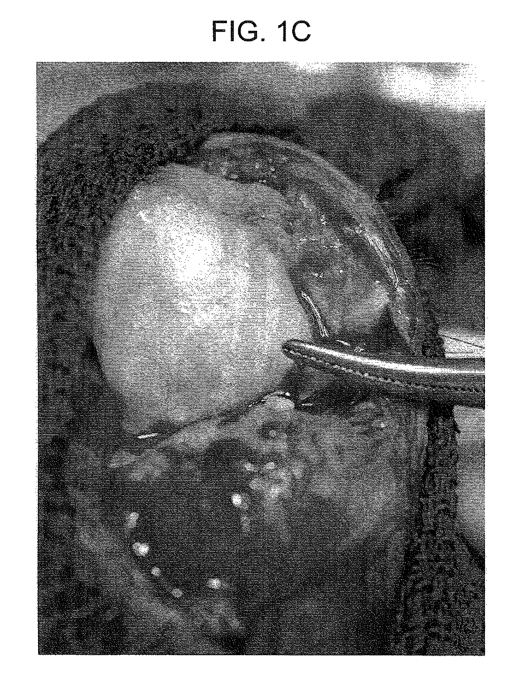
[0031]This example demonstrates the chondrogenic activity of the inventive composition in fully immunocompetant non-human primates. Full thickness cartilage defects measuring 10 min×10 mm were created in the medial condyles of the animals. The defects were densely packed with the cartilage composition according to the present invention and were compacted with a tamp. The animals were examined two, six, and sixteen weeks post transplantation and the joints were re-explored. FIGS. 1a-1c are photographs taken at two, six, and sixteen weeks, respectively. Specimens were also taken and were fixed in 10% formalin-Earle's balanced salt solutions. Paraffin sections were cut and stained with homotoxylin and eosin, PAS, Romanowski-Giemsa and Safranin-O stains. FIGS. 2a and 2b illustrate the specimens taken (100×) at six and sixteen weeks, respectively.

[0032]As seen in FIG. 1a, at two weeks post transplantation, granulation tissue is present in the center of the defect and new cartilage is pre...


PUM


Abstract

Disclosed is a method for regenerating articular cartilage in an animal comprising administering a therapeutically effective amount of a non-demineralized particulate articular cartilage having a distribution of particle sizes within the range of from about 60 microns to about 500 microns.

Description

CROSS-REFERENCE TO A RELATED APPLICATION[0001]This patent application is a divisional of copending U.S. patent application Ser. No. 10 / 793,939, filed Mar. 8, 2004, the disclosure of which is incorporated by reference.FIELD OF THE INVENTION[0002]This invention pertains to particulate cartilage compositions, processes for preparing particulate cartilage compositions and methods for regenerating articular cartilage.BACKGROUND OF THE INVENTION[0003]Articular cartilage of higher animals, including man, has very limited potential for repair. Following injuries to articular cartilage, the cartilage generally does not repair itself because of its very limited capacity for regeneration. Repair is dependent on the extent and the depth of injury and the surviving chondrocytes and normal articular cartilage matrix. In injuries involving subchondral bone, there is no regeneration of the cartilage, but there is typically enlargement and progression of the lesion with associated pain. The ultimate...

Claims


Application Information

Patent Timeline
Patent Type & Authority Patents(United States)
IPC IPC(8): A61K35/34A61F2/30A61L27/36
CPCA61L27/3612A61L27/3654A61L27/3691A61P19/02A61P19/04A61F2/30A61L27/36
Inventor MALININ, THEODORE
Owner VIVEX BIOLOGICS GRP INC
Learn More