Method for the diagnosis of lymphoproliferative diseases

a lymphoproliferative disease and lymphoid technology, applied in the field of new methods for diagnosis and treatment of lymphoid disease, can solve the problems of inability to definitively diagnose ctcl, difficult diagnosis of mf, and inability to detect m

Inactive Publication Date: 2010-09-28
VALIPHARMA
View PDF1 Cites 2 Cited by
  • Summary
  • Abstract
  • Description
  • Claims
  • Application Information

AI Technical Summary

Benefits of technology

[0011]We have now identified specific recurrent chromosomal breakpoints and / or translocations, which are associated with cutaneous T-cell lymphoma (CTCL), in chromosome 12, specifically in 12q14-12q24. We have also identified a gene involved in such chromosomal breakpoints and / or translocations. The identification of such specific chromosomal breakpoints and / or translocations and the gene and / or translocation, deletions or other defects of the gene allows the development of novel diagnostic and therapeutic methods for the detection and follow-up and treatment of lymphoproliferative diseases, such as CTCL.
[0013]Another object of the invention is to provide novel methods and means for the diagnosis of lymphoproliferative diseases, such as CTCL, such methods and means being specific and reliable.
[0014]Yet another object of the invention is to provide novel methods and means for the prediction of the progression of the disease and its transformation to an aggressive form in lymphoproliferative diseases, such as CTLC, such methods and means allowing a timely therapeutic intervention, which may be life-saving.
[0015]Still another object of the invention is to provide novel methods and means for the development of new guidelines for the initiation and follow-up of therapeutic interventions as well as for the development of new treatment modalities for lymphoproliferative diseases, such as CTCL, such methods and means prolonging the remission stage of the disease and introducing new possibilities for combating the disease and for the recovery of the patient.

Problems solved by technology

However, no means are available at present for a definite diagnosis of CTCL.
In particular, MF is difficult to diagnose in its early presentations due to its resemblance to eczema or mild psoriasis and unfortunately may thus remain undetected in time.
However, in the early phases of the disease these abnormalities have been non-clonal, which renders the detection non-specific.

Method used


Image

  • Method for the diagnosis of lymphoproliferative diseases
  • Method for the diagnosis of lymphoproliferative diseases
  • Method for the diagnosis of lymphoproliferative diseases

Examples


example 1

[0074]Identification of a Translocation in Chromosome 12

[0075]a) Cell Culture and Conventional Chromosome Preparations

[0076]Peripheral blood lymphocytes were isolated with Ficoll-Isopaque density centrifugation, washed with RPMI 1640 (Gibco BRL, Life Technologies) and cultured for 3 days in RPMI 1640, in the presence of 20% Fetal Bovine Serum (Gibco BRL), L-Glutamine (100× Liquid, used as 1×, Gibco BRL), antibiotics (Penicillin 10 000 IU / ml, Streptomycin 10 000 micrograms / ml, used as 1:100), and Phytohemagglutinin (PHA 10576-015, Gibco BRL, used as 1:100). After this the cells were treated with hypotonic KCl-solution, fixed with glacial acetic acid-methanol (1:3) and the cell suspension was dropped on objective slides to make conventional chromosome preparations.

[0077]b) MFISH-Method

[0078]The air-dried preparations were fixed with 0.1% paraformaldehyde and dried in ethanol series (70%, 85%, 100%). A probe mixture containing painting probes specific for each chromosome pair labelled ...

example 2

[0085]Characterization of the Translocation

[0086]The translocation was further defined with FISH by using locus-specific YAC-probes or BAC-probes (YAC-probes, YAC from CEPH, Fondation Jean Dausset, France; BAC-probes from Research Genetics, Groningen, tThe Netherlands). The YACs were grown in AHC broth and purified according to the instructions of Genomesystems (St. Louis, Mo., USA). The BACs were grown and purified according the instructions of Research Genetics (Groningen). Both YACs and BACs were labelled with FITC (Fluorescein-12-dUTP, NEN Life Science Products, Inc, Boston, Mass., USA), Alexa 488® or Alexa 594® (both Molecular probes, Leiden, The Netherlands) or biotin (Oncor Inc. Gaithersburg, Md., USA) using nick translation. The BACs were grown and purified according the instructions of Research Genetics (Groningen). For the YAC 803-C-2 and BAC RP11-359M6, see the chapter “Detailed description of the invention”.

[0087]The chromosomes involved were identified with centromere-s...

example 3

[0090]Identification of NAV3 Gene in the Translocation

[0091]For further identification of the translocation additional probes were used in FISH. YAC 855F7 (CEPH) was grown and purified and labelled with digoxigenin-11-dUTP (Roche) with nick-translation as described in Example 2. The hybridisation was performed as described above in Example 2 above and the hybridised probe was detected with sheep anti-digoxigenin-rhodamine antibodies (Roche, Mannheim).

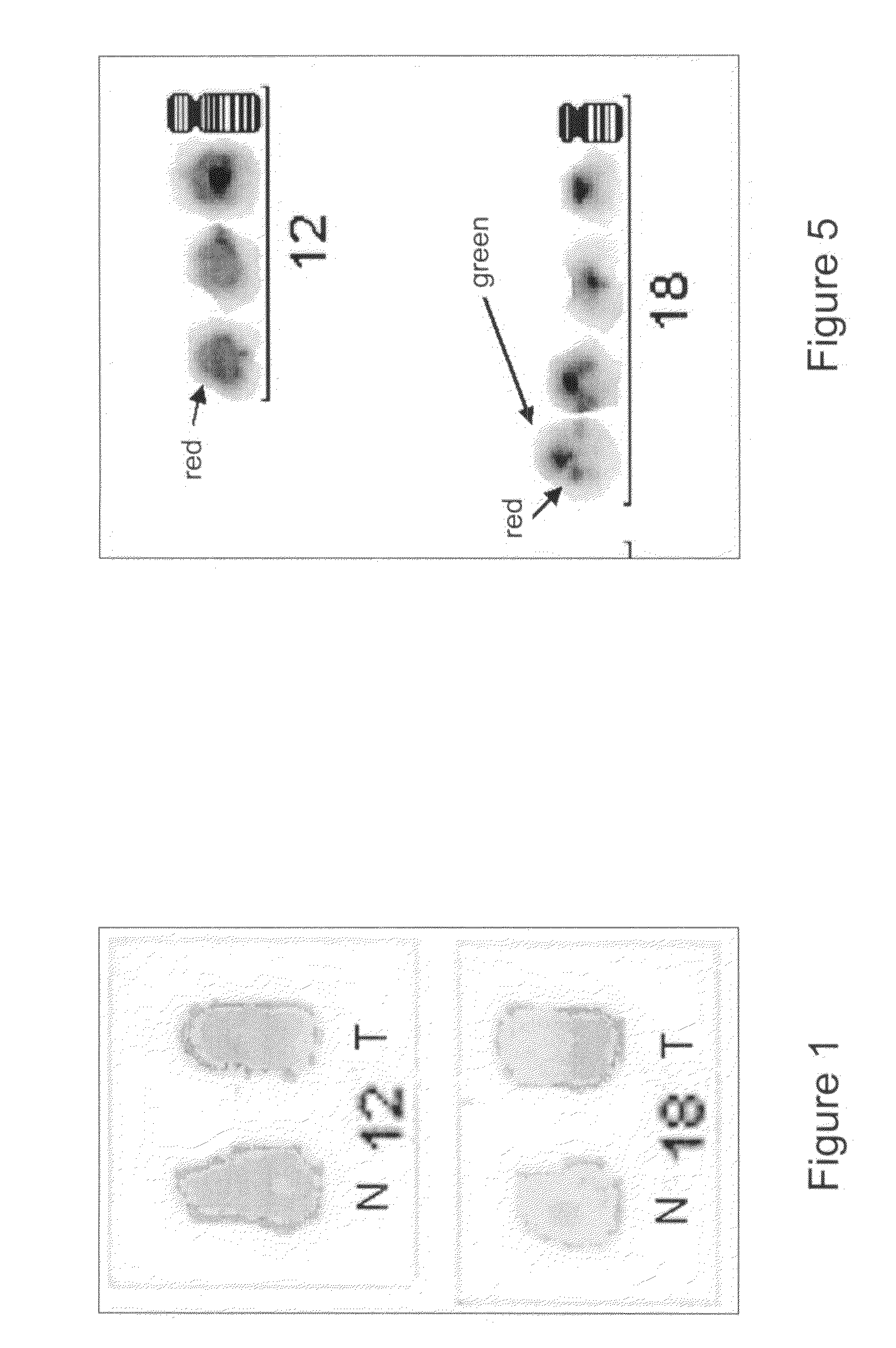
[0092]The hybridised probe was photographed and analysed with ultraviolet microscope and Isis 3-computer program (FIG. 8). In the sample from patient 3 suffering from SS, the YAC 855F7 divided between two chromosomes, 12q and 18q. In other hybridisations with further YACs the YACs situated in regions above YAC 855F7 (see FIG. 8, patient response block) remained on their respective places in the aberrant 12q, and the YACs below YAC 855F7 were found to be translocated from the aberrant 12q to the aberrant chromosome 18q. In samples from p...


PUM

PropertyMeasurementUnit
pHaaaaaaaaaa
pHaaaaaaaaaa
signal spectraaaaaaaaaaa

Abstract

The present invention relates to novel methods for the diagnosis and therapy of lymphoproliferative diseases. Specifically, the present invention relates to novel methods for the diagnosis and therapy taking advantage of the detection of chromosomal breakpoints in chromosome 12 and / or translocation of chromosomal material from chromosome 12, said chromosomal breakpoints and / or translocation(s) being associated with lymphoproliferative diseases, such as primary cutaneous T-cell lymphomas (CTCL). The present invention further relates to the use of neuron navigator 3 gene (NAV3) or an equivalent or functional fragment thereof involved in chromosomal breakpoints in chromosome 12 and / or translocations thereof, said gene and / or translocations thereof being associated with lymphoproliferative diseases, such as primary cutaneous T-cell lymphomas (CTCL), as a diagnostic and therapeutic agent. The present invention also relates to the development of therapy.

Description

[0001]This application is the US national phase of international application PCT / FI03 / 00061 filed 24 Jan. 2003, which designated the US and claims priority to FI Application No. 20020132 filed 24 Jan. 2002 and FI Application No. 20021617 filed 10 Sep. 2002. The entire contents of these applications are incorporated herein by reference.FIELD OF THE INVENTION[0002]The present invention relates to novel methods for the diagnosis and therapy of lymphoproliferative diseases. Specifically, the present invention relates to novel methods for the diagnosis and therapy taking advantage of the detection of chromosomal breakpoints in human chromosome 12 and / or translocation of chromosomal material from chromosome 12, said chromosomal breakpoints and / or translocation(s) being associated with lymphoproliferative diseases, such as primary cutaneous T-cell lymphomas (CTCL). The present invention further relates to the use of neuron navigator 3 gene (NAV3) or an equivalent or functional fragment the...

Claims


Application Information

Patent Timeline
Patent Type & Authority Patents(United States)
IPC IPC(8): C12Q1/68C12P19/34C07H21/02C07H21/04G01N33/50C12N15/09G01N21/78G01N33/53G01N33/566
CPCC12Q1/6886C12Q2600/156
Inventor RANKI, ANNAMARIKARENKO, LEENAKAHKONEN, MARKETTAKARHU, RITVAVISAKORPI, TAPIONEDOSZYTKO, BOGUSLAW
Owner VALIPHARMA
Why Eureka
Try Eureka