
Method and x-ray system for detecting position changes of a medical implant

a technology a position change, which is applied in the field of method and x-ray system for detecting the position change of a medical implant, can solve the problems of affecting the patient's life, affecting the patient's comfort, and the patient's stress, so as to reduce the time expenditure associated with post-operative examination, facilitate the necessary post-operative examination, and achieve the effect of very rapid implementation

Inactive Publication Date: 2010-02-09
SIEMENS HEALTHCARE GMBH
View PDF12 Cites 8 Cited by
  • Summary
  • Abstract
  • Description
  • Claims
  • Application Information

AI Technical Summary

Benefits of technology

The present invention provides a method for fast and automatic recognition of position changes of an implant using 2D x-ray exposures. The method uses markers that can be detected in the x-ray exposures and a threshold to determine if a position change has occurred. The method does not require a 3D model of the points to be calculated, reducing the dose of radiation and cost associated with post-operative examinations. The method can be implemented automatically without user intervention and can quantitatively determine the position change of the implant.

Problems solved by technology

Immediately after the implantation of artificial joints, the prosthesis can loosen in the course of time due to weakening of the materials used.
The method is tedious and additionally stresses the patient with the x-ray dose applied in the x-ray exposures.

Method used


Image

  • Method and x-ray system for detecting position changes of a medical implant
  • Method and x-ray system for detecting position changes of a medical implant
  • Method and x-ray system for detecting position changes of a medical implant

Examples


Embodiment Construction

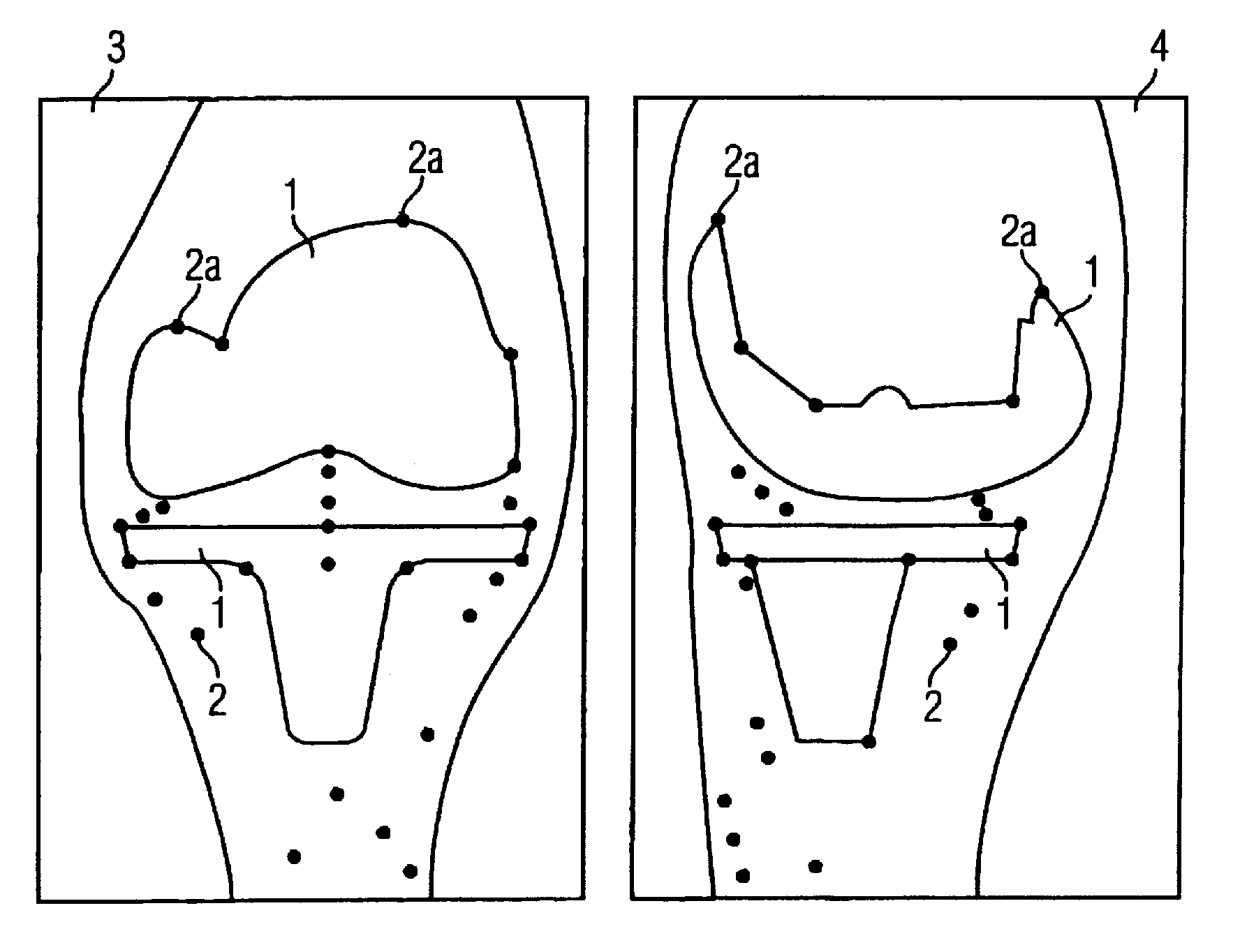
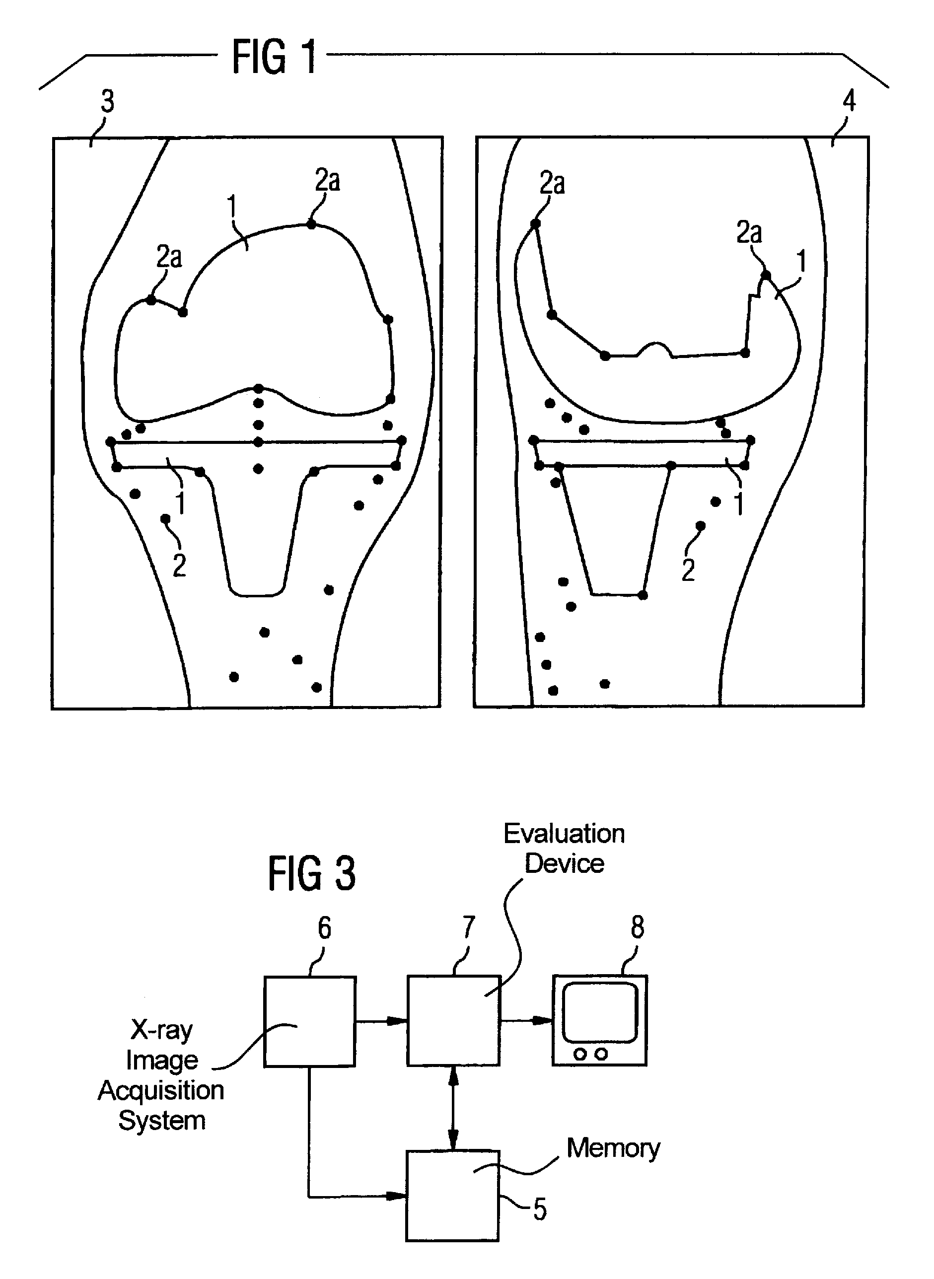
[0019]FIG. 1 shows an example two 2D x-ray exposures of a region containing an implant, in the present example a joint prosthetic 1 at the knee joint. The x-ray exposures were respectively obtained at different points in time from different directions, in the present example perpendicular to one another. In the x-ray exposures, respectively both components of the joint prosthetic 1 as well as metal spheres 2 (that are used as markers) placed in the bone are recognizable. Due to their placement in the bone, these metal spheres 2 do not change their position over the course of time. If a shifting of the joint prosthesis 1 occurs, this shifting can be recognized by a changed position of the prosthesis 1, or marked points 2a of the prosthesis 1, relative to the metal spheres 2 in the x-ray image.

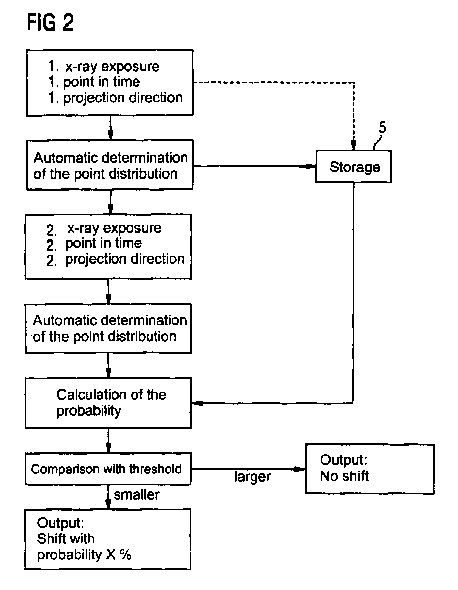
[0020]In the inventive method, the distribution of the metal spheres 2 as well as marked points 2a are automatically determined both in the first x-ray exposure 3 and in the second x-ray exposur...


PUM


Abstract

In a method and apparatus to detect position changes of an implant x-ray detectable markers are arranged in the environment of the implant, and 2D x-ray exposures are obtained at temporal intervals of a region containing the implant in which a distribution of the markers as well as of marked points of the implant are visible in each 2D x-ray exposure. A first of the 2D x-ray exposures is obtained at a first point in time from one projection direction, and a second of the 2D x-ray exposures is obtained at a second point in time from another projection direction. The distribution of the markers and marked points is determined in the first and second 2D x-ray exposure by an evaluation device, and from the distributions a degree of probability is calculated that the distribution of the first 2D x-ray exposure and the distribution of the second 2D x-ray exposure are projections of the same three-dimensional distribution of markers and marked points. An automated detection of position changes by implants with reduced x-ray exposure for the patient is facilitated.

Description

BACKGROUND OF THE INVENTION[0001]1. Field of the Invention[0002]The present invention concerns a method to detect position changes of a medical wherein x-ray detectable markers are arranged in the environment of the implant, and wherein a positional change of the implant is checked using 2D x-ray exposures at temporal intervals of a region containing the implant, using a distribution of the markers in each 2D x-ray exposure, with a first of the 2D x-ray exposures being obtained at a first point in time from one projection direction, and a second of the 2D x-ray exposures being obtained at a second point in time from another projection direction. The invention also concerns an x-ray system fashioned for implementation of the method.[0003]2. Description of the Prior Art[0004]Supervision of the change of the position of implants, in particular the position of implanted prostheses, is an important post-operative measure in order to be able to undertake timely corrections and to prevent ...

Claims


Application Information

Patent Timeline
Patent Type & Authority Patents(United States)
IPC IPC(8): A61B5/05A61B6/12
CPCA61B6/12A61F2002/3008A61F2002/4658A61F2002/48A61F2250/0098A61F2/48
Inventor GEORG, CHRISTIANHORNEGGER, JOACHIMKLOSE, KLAUS JOCHENWELKER, VOLKMAR
Owner SIEMENS HEALTHCARE GMBH
Learn More