Methods and devices for performing cardiac valve repair
a technology for cardiac valves and valves, applied in medical science, surgery, surgical forceps, etc., can solve the problems of affecting the proper functioning of one or more cardiac valves, prolapse and regurgitation, and obstruction of blood flow, so as to prevent valvular regurgitation
- Summary
- Abstract
- Description
- Claims
- Application Information
AI Technical Summary
Benefits of technology
Problems solved by technology
Method used
Image


Examples
Embodiment Construction
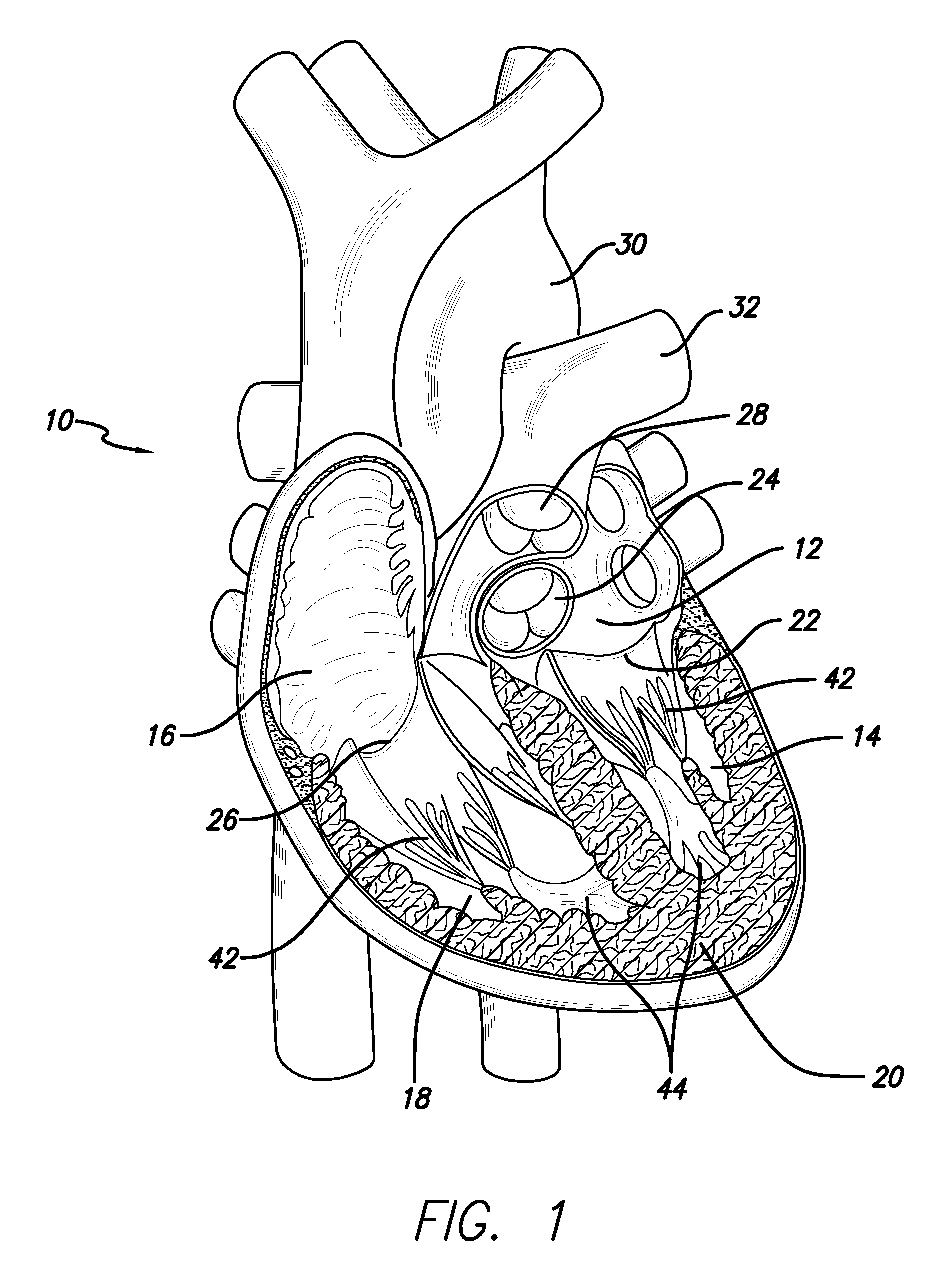
[0037]The methods disclosed herein set forth minimally invasive procedures that both simplify and expedite cardiac valve repairs. Unlike conventional protocols, the methods herein presented do not involve invasive surgical procedures and do not require cardioplegic arrest and / or lengthy cardiopulmonary bypass. Rather the methods of the invention may be performed while the heart is still beating, thereby reducing if not entirely avoiding pump and cross-clamp time, minimizing pain and trauma, and promoting rapid recovery.
[0038]In order to perform a cardiac valve repair of the invention, one or more chambers of the heart must be accessed. Rather than requiring open heart surgery as a means of accessing a chamber of the heart, which necessitates the stopping and bypassing of the heart by an extracorporeal circulation device, the methods of the invention are performed in a minimally invasive manner. Using small incisions, specialized instruments, and / or video / audio-scopic assistance (e.g...
PUM
 Abstract
Description
Claims
Application Information
 - R&D
- Intellectual Property
- Life Sciences
- Materials
- Tech Scout
- Unparalleled Data Quality
- Higher Quality Content
- 60% Fewer Hallucinations
Browse by: Latest US Patents, China's latest patents, Technical Efficacy Thesaurus, Application Domain, Technology Topic, Popular Technical Reports.
© 2025 PatSnap. All rights reserved.Legal|Privacy policy|Modern Slavery Act Transparency Statement|Sitemap|About US| Contact US: help@patsnap.com