
Specific binding proteins and uses thereof

a technology of specific binding proteins and binding members, which is applied in the field of specific binding members, can solve the problems of limited use of these antibodies, poor prognosis of expression of egfr, and limitations of the range of applicability and efficacy reflected above, and achieves significant in vivo anti-tumor activity.

Inactive Publication Date: 2009-09-15
LUDWIG INST FOR CANCER RES LTD
View PDF3 Cites 98 Cited by
  • Summary
  • Abstract
  • Description
  • Claims
  • Application Information

AI Technical Summary

Benefits of technology

[0014]The unique specificity of mAb 806, whereby mAb 806 binds to the de2-7 EGFR and amplified EGFR but not the normal, wild type EGFR, provides diagnostic and therapeutic uses to identify, characterize and target a number of tumor types, for example, head and neck, breast, or prostate tumors and glioma, without the problems associated with normal tissue uptake that may be seen with previously known EGFR antibodies.
[0031]The present invention naturally contemplates several means for preparation of the antibodies and active fragments thereof, including as illustrated herein known recombinant techniques, and the invention is accordingly intended to cover such synthetic or chimeric antibody preparations within its scope. The isolation of the cDNA and amino acid sequences disclosed herein facilitates the reproduction of the antibody of the present invention by such recombinant techniques, and accordingly, the invention extends to expression vectors prepared from the disclosed DNA sequences for expression in host systems by recombinant DNA techniques, and to the resulting transformed hosts.

Problems solved by technology

Moreover, expression of the EGFR is associated with poor prognosis in a number of tumor types including stomach, colon, urinary bladder, breast, prostate, endometrium, kidney and brain (e.g., glioma).
The use of these antibodies, however, may be limited by uptake in organs that have high endogenous levels of EGFR such as the liver and skin (18, 19).
Thus, while the extant evidence of activity of EGFR antibodies is encouraging, the observed limitations on range of applicability and efficacy reflected above remain.

Method used


Image

  • Specific binding proteins and uses thereof
  • Specific binding proteins and uses thereof
  • Specific binding proteins and uses thereof

Examples


example 1

Isolation of Antibodies

Materials

Cell Lines

[0294]For immunization and specificity analyses, several cell lines, native or transfected with either the normal, wild type or “wtEGFR” gene or the ΔEGFR gene carrying the Δ2-7 deletion mutation were used: Murine fibroblast cell line NR6, NR6ΔEGFR (transfected with ΔEGFR) and NR6wtEGFR (transfected with wtEGFR), human glioblastoma cell line U87MG (expressing low levels of endogenous wtEGFR), U87MGwtEGFR (transfected with wtEGFR), U87MGΔEGFR (transfected with ΔEGFR), and human squamous cell carcinoma cell line A431 (expressing high levels of wtEGFR)[38]. Cell lines and transfections were described previously (Nishikawa R., et al. (1994) Proc. Natl. Acad. Sci. 91(16):7727-7731).

[0295]The U87MG astrocytoma cell line (20), which endogenously expresses low levels of the wt EGFR, was infected with a retrovirus containing the de2-7 EGFR to produce the U87MG.Δ2-7 cell line (10). The transfected cell line U87MG.wtEGFR was produced as described in Na...

example 2

Binding of Antibodies to Cell Lines by FACS

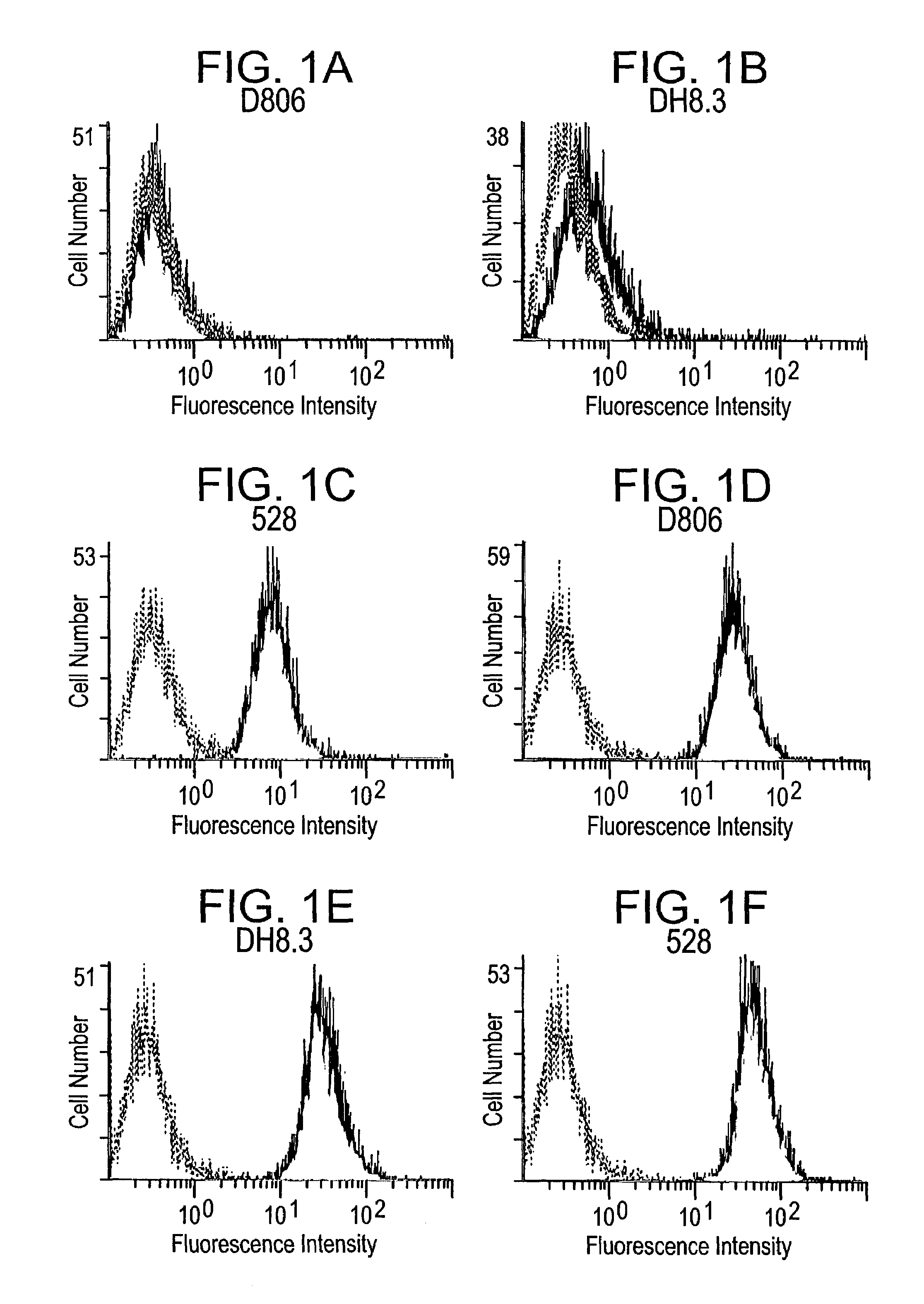
[0304]In order to determine the specificity of mAb 806, its binding to U87MG, U87MG.Δ2-7 and U87MG.wtEGFR cells was analyzed by flow activated cell sorting (FACS). Briefly, cells were labelled with the relevant antibody (10 μg / ml) followed by fluorescein-conjugated goat anti-mouse IgG (1:1100 dilution; Calbiochem San Diego, USA). FACS data was obtained on a Coulter Epics Elite ESP by observing a minimum of 5,000 events and analysed using EXPO (version 2) for Windows. An irrelevant IgG2b was included as an isotype control for mAb 806 and the 528 antibody was included as it recognizes both the de2-7 and wt EGFR.

[0305]Only the 528 antibody was able to stain the parental U87MG cell line (FIG. 1) consistent with previous reports demonstrating that these cells express the wt EGFR (Nishikawa et al, 1994). MAb 806 and DH8.3 had binding levels similar to the control antibody, clearly demonstrating that they are unable to bind the wt receptor (FIG. 1...

example 3

Binding of Antibodies in Assays

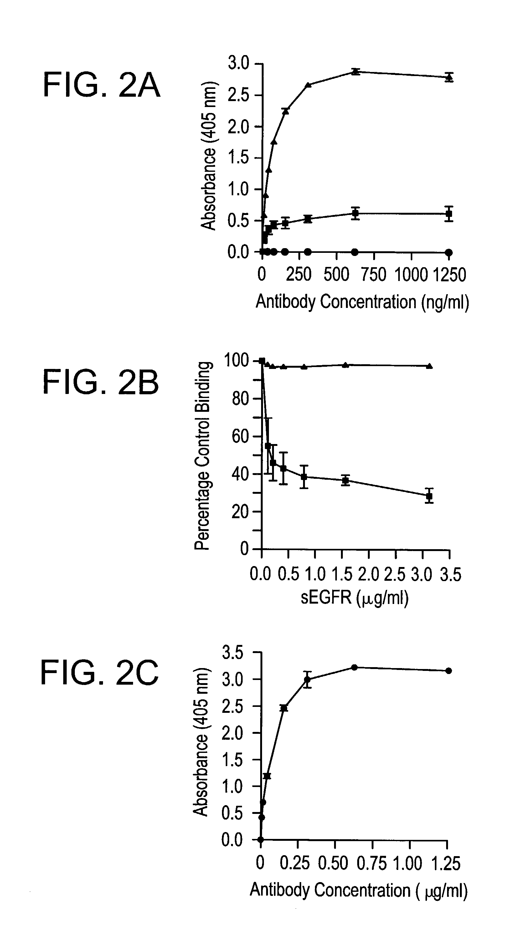
[0307]To further characterize the specificity of mAb 806 and the DH8.3 antibody, their binding was examined by ELISA. Two types of ELISA were used to determine the specificity of the antibodies. In the first assay, plates were coated with sEGFR (10 μg / ml in 0.1 M carbonate buffer pH 9.2) for 2 h and then blocked with 2% human serum albumin (HSA) in PBS. sEGFR is the recombinant extracellular domain (amino acids 1-621) of the wild type EGFR), and was produced as previously described (22). Antibodies were added to wells in triplicate at increasing concentration in 2% HSA in phosphate-buffered saline (PBS). Bound antibody was detected by horseradish peroxidase conjugated sheep anti-mouse IgG (Silenus, Melbourne, Australia) using ABTS (Sigma, Sydney, Australia) as a substrate and the absorbance measured at 405 nm.

[0308]Both mAb 806 and the 528 antibody displayed dose-dependent and saturating binding curves to immobilized wild type sEGFR (FIG. 2A). As the u...


PUM


Abstract

The invention relates to specific binding members, particularly antibodies and active fragments thereof, which recognize an aberrant post-translationally modified, particularly an aberrant glycosylated form of the EGFR. The binding members, particularly antibodies and fragments thereof, of the invention do not bind to EGFR on normal cells in the absence of amplification of the wild-type gene and are capable of binding the de2-7 EGFR at an epitope which is distinct from the junctional peptide. Antibodies of this type are exemplified by the novel antibody 806 whose VH and VL sequences are illustrated as SEQ ID NOs: 2 and 4 and chimeric antibodies thereof as exemplified by ch806.

Description

CROSS REFERENCE TO RELATED APPLICATIONS[0001]The present application claims priority from U.S. Provisional Application Ser. No. 60 / 290,410, filed May 11, 2001, Ser. No. 60 / 326,019, filed Sep. 28, 2001, and Ser. No. 60 / 342,258, filed Dec. 21, 2001, each of which is incorporated by reference herein in its entirety.FIELD OF THE INVENTION[0002]The present invention relates to specific binding members, particularly antibodies and fragments thereof, which bind to amplified epidermal growth factor receptor (EGFR) and to the de2-7 EGFR truncation of the EGFR. In particular, the epitope recognized by the specific binding members, particularly antibodies and fragments thereof, is enhanced or evident upon aberrant post-translational modification. These specific binding members are useful in the diagnosis and treatment of cancer. The binding members of the present invention may also be used in therapy in combination with chemotherapeutics or anti-cancer agents and / or with other antibodies or fr...

Claims


Application Information

Patent Timeline
Patent Type & Authority Patents(United States)
IPC IPC(8): C07K16/28A61K39/00G01N33/50A61K39/395A61K45/00A61K51/00A61P35/00C07K16/32C07K16/46C07K19/00C12N1/19C12N1/21C12N5/10C12N15/09C12P21/08G01N33/15G01N33/53G01N33/566
CPCA61K39/39541C07K16/2863A61K45/06A61K39/395A61K2039/505C07K2317/732C07K2317/734C07K2317/34C07K2317/77A61K2300/00A61P35/00
Inventor OLD, LLOYD J.JOHNS, TERRANCE GRANTPANOUSISSCOTT, ANDREW MARKRENNER, CHRISTOPHRITTER, GERDJUNGBLUTH, ACHIMSTOCKERT, ELISABETHCOLLINS, PETERCAVENEE, WEBSTER K.HUANG, HUEI-JEN SUBURGESS, ANTONY WILKSNICE, EDOUARD COLLINS
Owner LUDWIG INST FOR CANCER RES LTD
Learn More