
Real time brachytherapy spatial registration and visualization system

Inactive Publication Date: 2007-04-10
IMPAC MEDICAL SYST
View PDF171 Cites 51 Cited by
  • Summary
  • Abstract
  • Description
  • Claims
  • Application Information

AI Technical Summary

Benefits of technology

[0018]It is yet a further object of the invention to provide an automated method and system for loading of radioactive therapeutic treatment

Problems solved by technology

New minimally invasive surgical procedures are most often optically guided, but such optical guidance methods do not permit visualization and guidance of instruments or probes within (inside) the target tissue or organ.
However, the method is expensive, not truly real-time, and is limited in application.
These methods suffer from inaccuracy of localizing the target for any one particular individual and potential real time changes of relative orientation and position of target tissue, normal tissue, and radiation therapy devices.
There was significant dose inhomogeneity due to the nonuniformity of seed placement, leading to underdosing of portions of the prostate gland and significant complications due to overdosing of adjacent healthy tissue structures.
The poor results for local control and normal tissue complication were attributed to the doctor's inability to visualize and hence control where the radioactive seeds were actually being deposited inside the patient.
Even with this technique, significant imprecisions in seed placement are observed.
Due to the proximity of the prostate to the rectum and bladder, incorrect seed placement may lead to serious overdosing of these structures and late complications.
This is a time-consuming process; and it is very difficult to achieve optimal placement.
Invariably, the catheters deflect angularly as they are inserted, and their positions are difficult to determine by two-dimensional ultrasound.
These above described prior art systems suffer from inherent inaccuracy, the inability to correct the positioning of the radioactive seeds without repeated withdrawal and reinsertion of seeds into the prostate and are not real time manipulations of the therapeutic medium.
Consequently, the catheter position and seed position may be at an undesired position relative to the presumed assessment phase location.

Method used


Image

  • Real time brachytherapy spatial registration and visualization system
  • Real time brachytherapy spatial registration and visualization system
  • Real time brachytherapy spatial registration and visualization system

Examples


Embodiment Construction

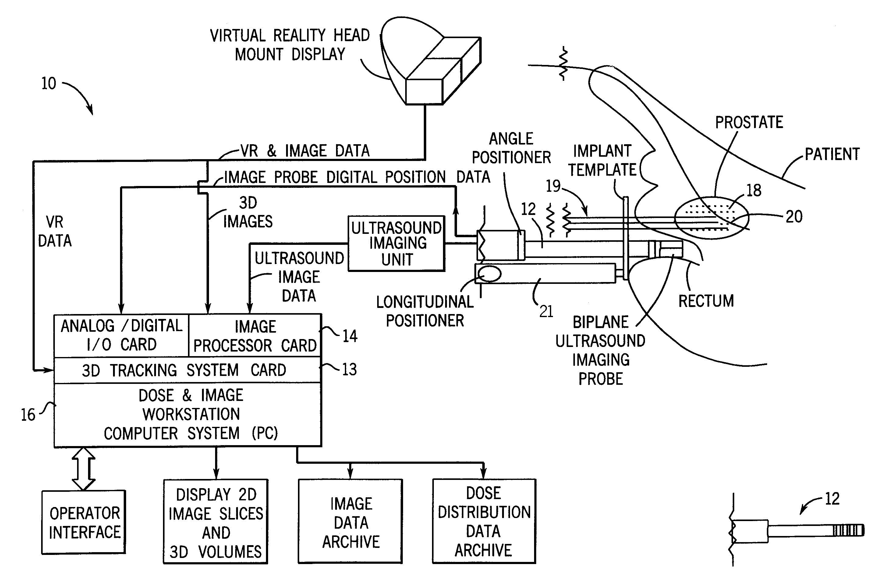
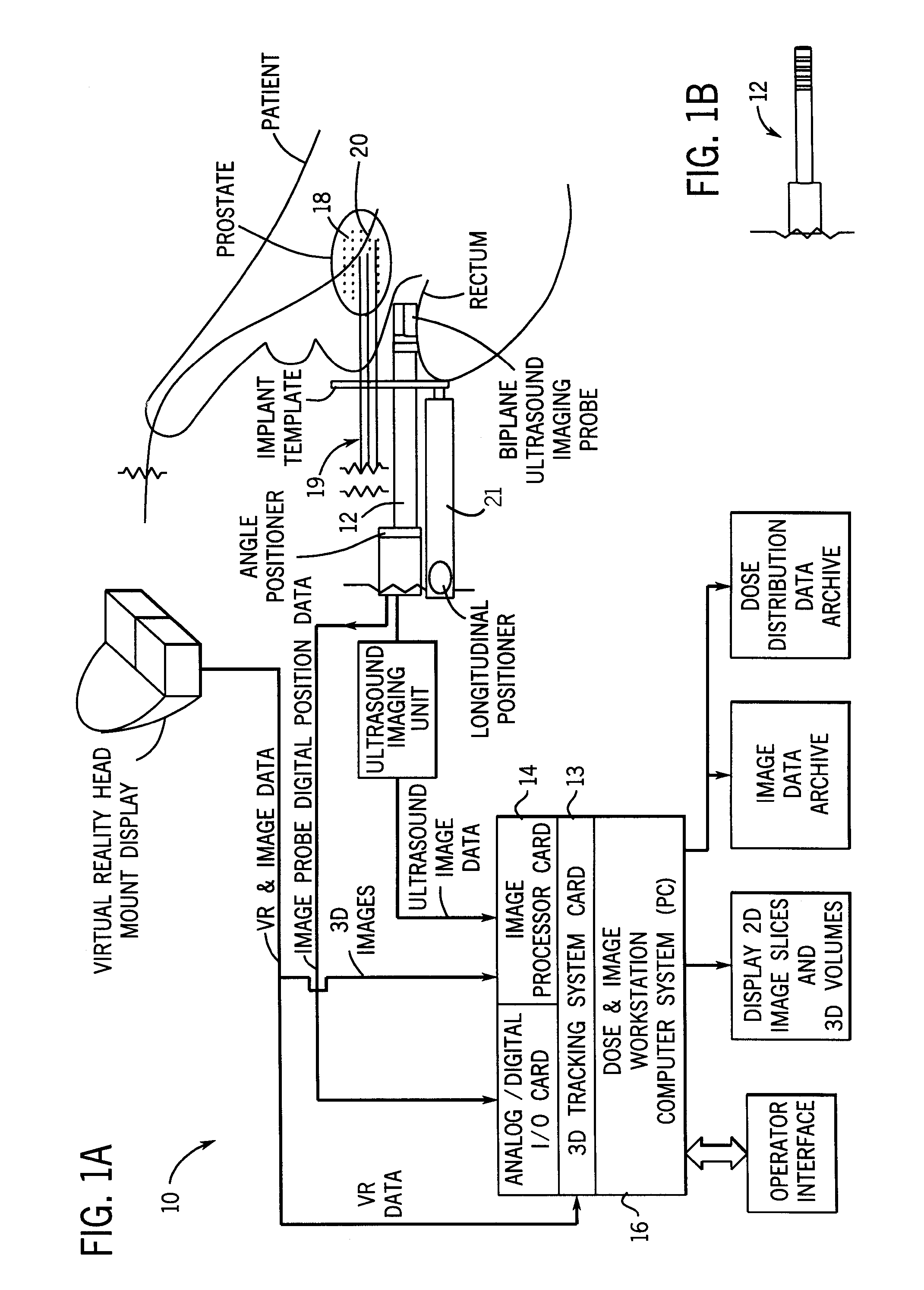
[0034]A system 10 constructed in accordance with an example of the invention is illustrated generally in FIG. 1A. A three-dimensional probe 12 accumulates image data from a treatment region or organ of a patient, image data is processed using a three-dimensional imaging card 14. The probe 12 preferably is an ultrasound device but can be any other rapid imaging technology, such as rapid CT or MR. A conventional personal computer 16 having a monitor can be used to operate on the image data from the imaging card 14 using conventional software and hardware tools to be described in more detail hereinafter. Radioactive seeds 18 are provided for insertion using any one of a variety of conventional means for inserting devices or articles into the human body, such as insertion devices 19, which may be either needles or stiff catheters. The three-dimensional ultrasound probe 12, therefore, provides an image signal to the computer 16 and a virtual reality interface card 13 coupled to the imagi...


PUM


Abstract

A method and system for monitoring loading of radiation into an insertion device for use in a radiation therapy treatment to determine whether the treatment will be in agreement with a radiation therapy plan. In a preferred embodiment, optical sensors and radiation sensors are used to monitor loading of radioactive seeds and spacers into a needle as part of a prostate brachytherapy treatment.

Description

CROSS-REFERENCE AND PRIORITY CLAIMS TO RELATED APPLICATIONS[0001]This patent application is a continuation of U.S. application Ser. No. 09 / 573,415 filed on May 18, 2000, now U.S. Pat. No. 6,512,942, which is a continuation of U.S. application Ser. No. 09 / 087,453 filed on May 29, 1998, now U.S. Pat. No. 6,129,670, which is a continuation-in-part of U.S. application Ser. No. 08 / 977,362 filed on Nov. 24, 1997, now U.S. Pat. No. 6,256,529.CROSS-REFERENCE TO COMPUTER PROGRAM LISTING ON COMPACT DISC[0002]This patent application includes a Computer Program Appendix on compact disc.BACKGROUND AND SUMMARY OF THE INVENTION[0003]The present invention is directed in general to an improved method and apparatus for carrying out minimally invasive treatments of the human body by virtual reality visualization of the treatment area. More particularly the invention is concerned with use of an apparatus and method for providing real time images of a human anatomy undergoing treatment along with rapid ...

Claims


Application Information

Patent Timeline
IPC IPC(8): A61N5/00A61B5/055A61B6/00A61B8/12A61B17/00A61B17/34A61B18/22A61B19/00A61M36/04A61N5/10
CPCA61B8/12A61B8/42A61B8/4209A61B8/5238A61B19/52A61B19/5244A61M37/0069A61N5/1001A61N5/1002A61N5/1007A61N5/103A61N5/1048A61N5/1049A61B5/055A61B18/22A61B19/50A61B2017/00274A61B2017/3411A61B2018/00547A61B2019/5276A61B2019/5278A61B2019/5291A61B2019/566A61N5/1027A61N2005/1011A61N2005/1012A61B90/36A61B2090/3782A61B2034/256A61B34/20A61B34/10A61B2090/378A61B2090/365A61N5/00
Inventor BURDETTE, EVERETTE C.KOMANDINA, BRUCE M.
Owner IMPAC MEDICAL SYST
Learn More