Eureka AIR delivers breakthrough ideas for toughest innovation challenges, trusted by R&D personnel around the world.

Time share digital integration method and apparatus for processing X-ray images

a digital integration and image technology, applied in the field of time-share digital integration methods and apparatuses for processing x-ray images, can solve the problems of short and unnecessarily large and complex electronic signal processing circuitry, etc., to achieve the effect of increasing the data transfer rate of detector array boards, increasing the cost of the board, and increasing the time required for outputting image data

Inactive Publication Date: 2006-11-21
LE KHAI MINH
View PDF6 Cites 65 Cited by
  • Summary
  • Abstract
  • Description
  • Claims
  • Application Information

AI Technical Summary

Benefits of technology

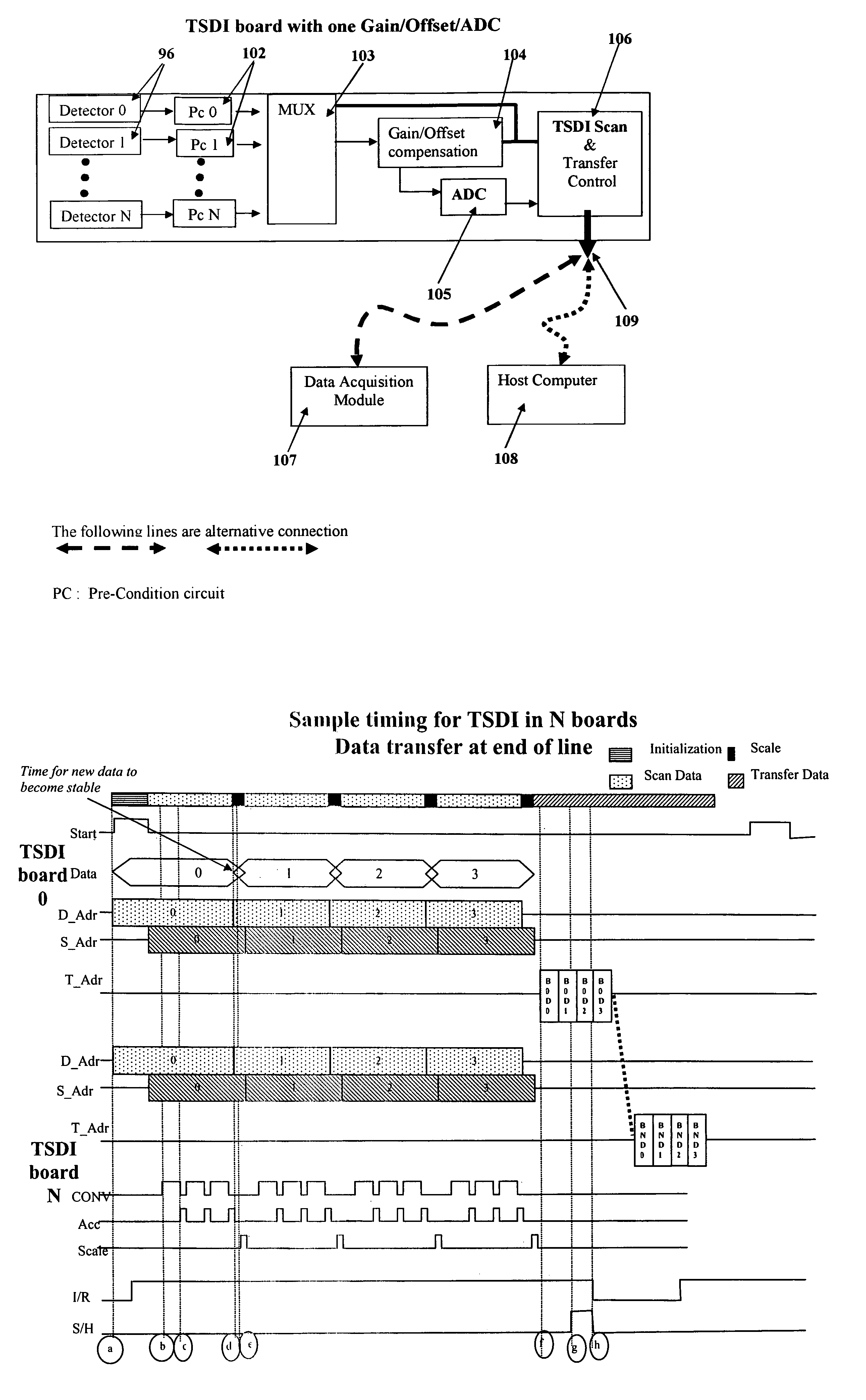
[0026]Another object of the invention is to provide an X-ray-ray detector array board in which signals output from a plurality of X-ray radiation detectors are each sampled during each of a plurality of scan lines of an object field raster sampled repetitively and periodically P times and accumulated in a digital integrator to thereby increase signal-to-noise ratio of a board output signal by a technique called Time Share Digital Integration (TSDI).
[0045]Digital values representing image information obtained from each detector must ultimately be output from a detector array board to a host computer, either directly or Into a digital acquisition module, where data from multiple image slices of an object obtained on one or more detector array boards is concatenated to form a complete electronic raster image of the object. The electronic image is used by the host computer or other external equipment to produce a visual image and / or be processed by pattern recognition software to detect guns, explosives, etc. Since a typical system according to the present invention has a thousand or more detectors, the time required for outputting image data can become significant. Although various high speed data transfer techniques such as LVDS (Low Voltage Differential Signal) are available, implementing such techniques on each detector array board can significantly increase the cost of the board because of the number of required components and their cost. Preferably, therefore, in accordance with one aspect of the present invention, detector array board data transfer rates are increased by the following method.

Problems solved by technology

The exposure time is kept as short as possible because X-radiation has a cumulative destructive affect on biological cells.
Thus, because simultaneous parallel processing of 1024 detector signal channels would required unnecessarily large and complex electronic signal processing circuitry, most signal processing circuitry currently used on scanning X-ray radiography systems sequentially samples the outputs of a vertical detector array.
A shorter time constant will result in a sharper image but more photon statistic noise.
Scanning X-ray radiography systems using a single ADC require transmission of analog signals through a cable / bus which may be many feet long; therefore, such systems are subject to noise interference from the environment.
ADC on each board increases the cost of each board, such boards often are provided with true integrators in place of pseudo-integrators.
The cost and complexity of each second type, true integrator detector array board is substantially greater than that of the first-type, pseudo-integrator board described above.
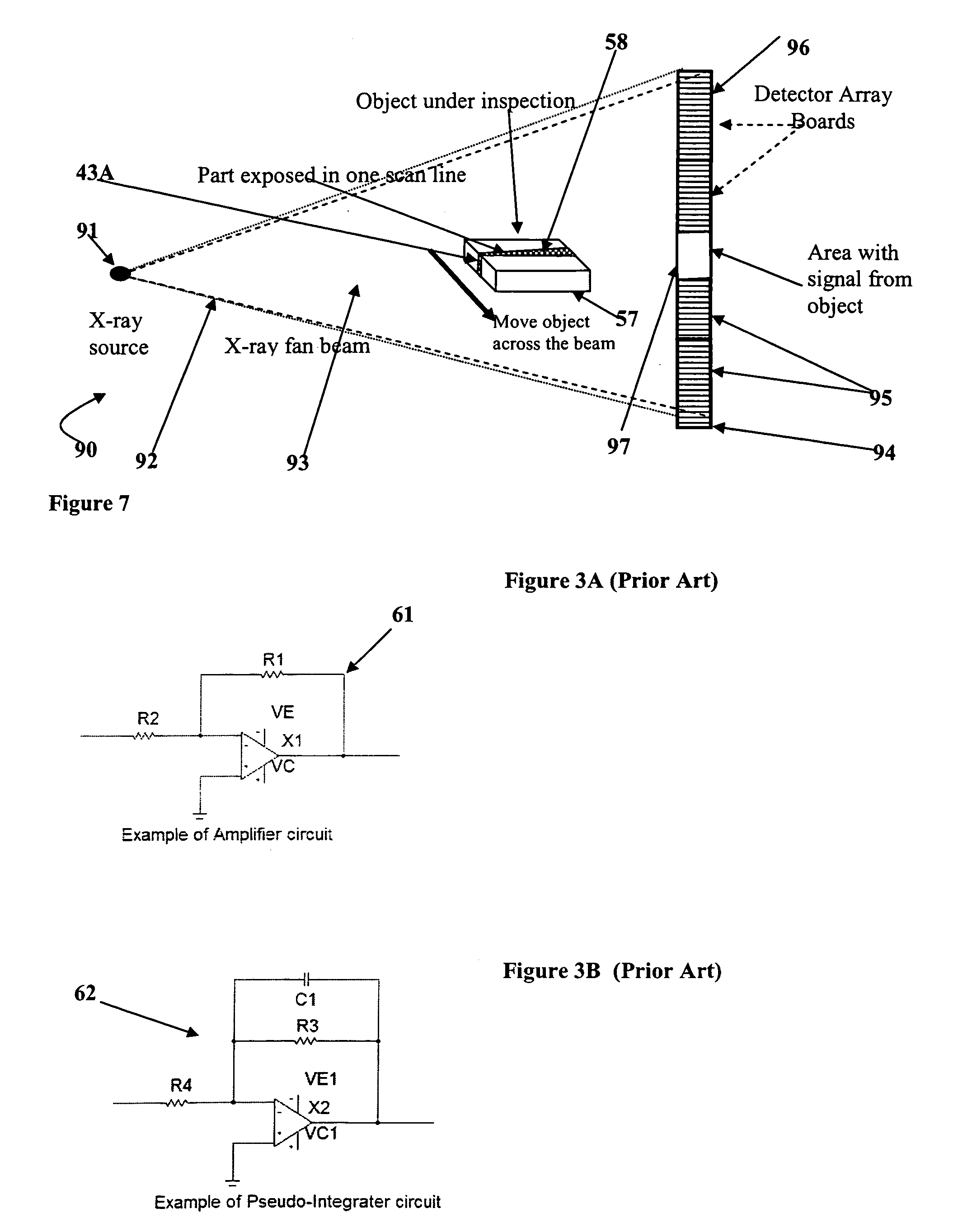
In this implementation, selecting a time constant for the pseudo-integrator that is relatively long will result in low photon-statistic noise, but if the time constant is too long, electronic images derived from the signals will be blurred.
A shorter pseudo-integrator time constant will result in a sharper image, but if made too short, variations in image brightness caused by photon statistic variations will become noticeable, particularly in low signal areas, and result in a grainy image.
In the second type, true-integrator detector array board described above, a practical limitation is imposed on board performance by a limitation in the amount of charge that each detector channel integrator circuit can hold.
Using the same integrator circuits in a system with high power X-ray-ray sources or longer integration times can cause the charge storage capacitor of an integrator circuit to reach a maximum, saturated level while the detector output signal is still increasing, thus causing image information to be lost.
As mentioned above, the third type detector array board implementation which has a separate ADC for each detector channel avoids the limitations of the pseudo-integrator and true integrator boards described above, but is costly.

Method used


Image

  • Time share digital integration method and apparatus for processing X-ray images
  • Time share digital integration method and apparatus for processing X-ray images
  • Time share digital integration method and apparatus for processing X-ray images

Examples


Embodiment Construction

[0066]The structure and function of a Time-Share Digital Integration (TSDI) method and apparatus according to the present invention, and certain novel and advantageous improvements in X-ray radiography systems afforded by the method and apparatus, may be best understood by first reviewing the construction and function of some prior art X-ray radiography systems.

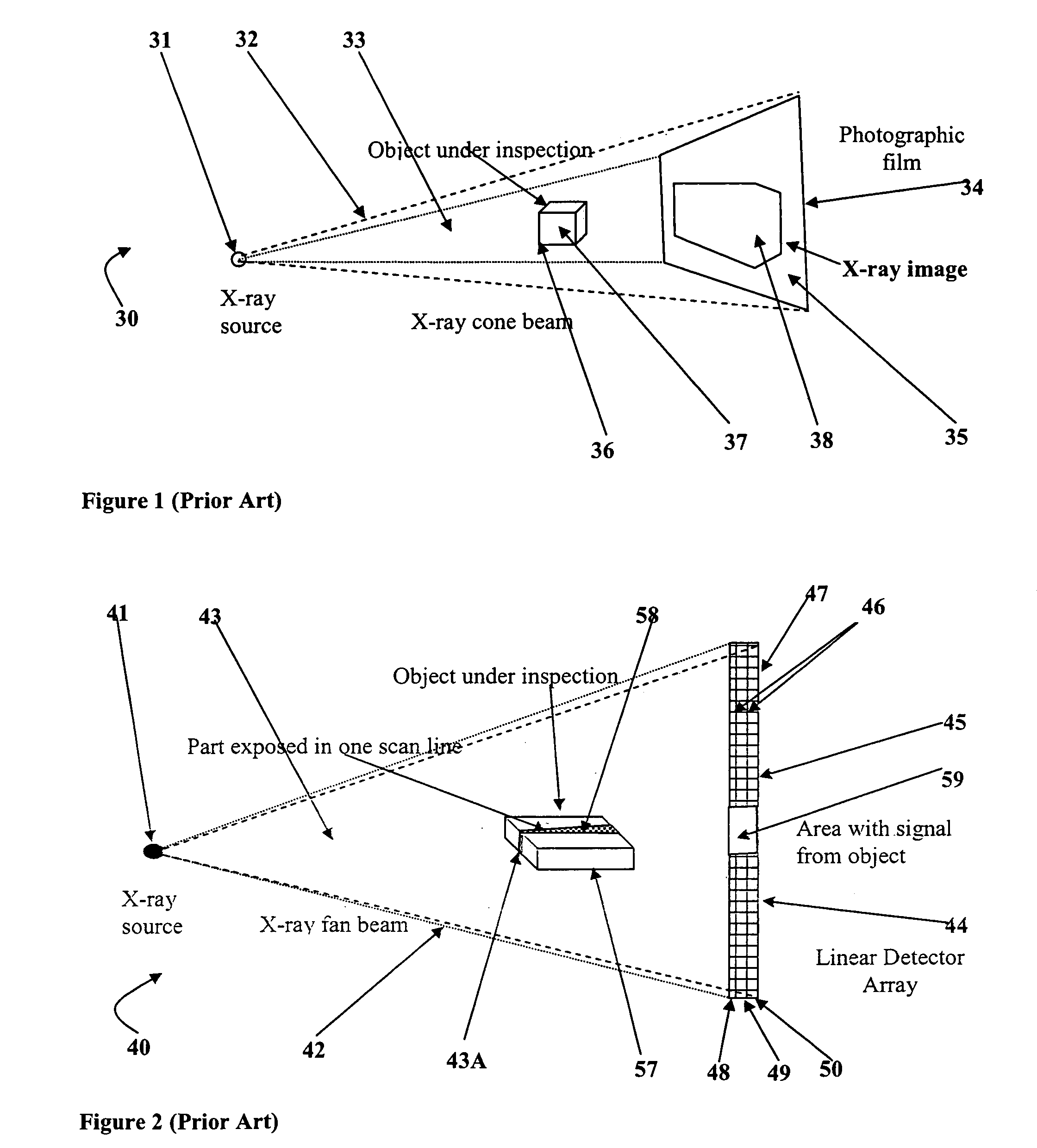
[0067]FIG. 1, shows a basic prior art X-ray transmission radiography system for obtaining images of internal features of human bodies, cargo containers, luggage or the like. As shown in FIG. 1, a typical prior art X-ray radiography system 30 includes a source of X-ray radiation 31 such as an X-ray tube, which produces a generally conically-shaped beam of X-ray radiation 32 that irradiates or illuminates an object space 33 and an image plane 34 located on a side of the object space opposite that of X-ray radiation source 31. At the image plane 34 is located a device for detecting X-ray radiation such as a fluorescent screen, p...


PUM


Abstract

A method and apparatus for processing signals output from an array of X-ray radiation detectors which receive X-ray radiation emanating from an object irradiated by a source beam of X-ray radiation, to thereby produce an visual image of internal object features, utilizes repetitive sampling P times and digital accumulation of signals output from each detector to increase signal-to-noise ratio of the images by the factor 1 / ✓P. In a basic embodiment, each of the N detectors in a linear detector array is sampled P times. In another embodiment, artifacts resulting from decay of detector pre-amplifier output voltages on capacitors of low-pass filter circuits, referred to as pseudo-integrators, are reduced by sequentially sampling and accumulating each of N detector amplifier output voltages P times in turn, and repeating the sampling and accumulating sequence Q times, thereby reducing variations in the P×Q accumulated values between the first and last detector channels of an N detector array.

Description

BACKGROUND OF THE INVENTION[0001]A. Field of the Invention[0002]The present invention relates to methods and apparatus which employ X-ray or gamma ray radiography to obtain images of internal features of a human body, or objects within luggage or other such containers. More particularly, the invention relates to improved signal processing methods and apparatus for converting signals output from an array of X-ray radiation detectors which receive X-radiation emanating from an object irradiated by a source beam of X-ray radiation, to visually discernible images of internal features of the object.[0003]B. Description of Background Art[0004]Electromagnetic radiation of wavelengths substantially shorter than visible light, specifically X-rays and gamma rays having wavelengths less than about 0.1 nm, are routinely used to obtain visually discernible images of internal or sub-surface features of an object, by a method referred to as radiography. For example, transmission X-radiography has ...

Claims


Application Information

Patent Timeline
IPC IPC(8): H05G1/64
CPCG01V5/0008G01V5/20
Inventor LE, KHAI MINH
Owner LE KHAI MINH
Why Eureka
Eureka Blog
Learn More