
Segmentation driven image noise reduction filter

a segmentation driven, filter technology, applied in image enhancement, image analysis, visual presentation, etc., can solve the problems of difficult to determine which of these parameters, or which combination of parameters, may be adjusted to provide optimal image presentation, and known signal processing systems for enhancing discrete pixel images suffer from certain drawbacks, etc., to achieve computational efficiency, maintain image quality, and enhance discrete pixel images

Inactive Publication Date: 2005-12-06
GE MEDICAL SYST GLOBAL TECH CO LLC
View PDF8 Cites 9 Cited by
  • Summary
  • Abstract
  • Description
  • Claims
  • Application Information

AI Technical Summary

Benefits of technology

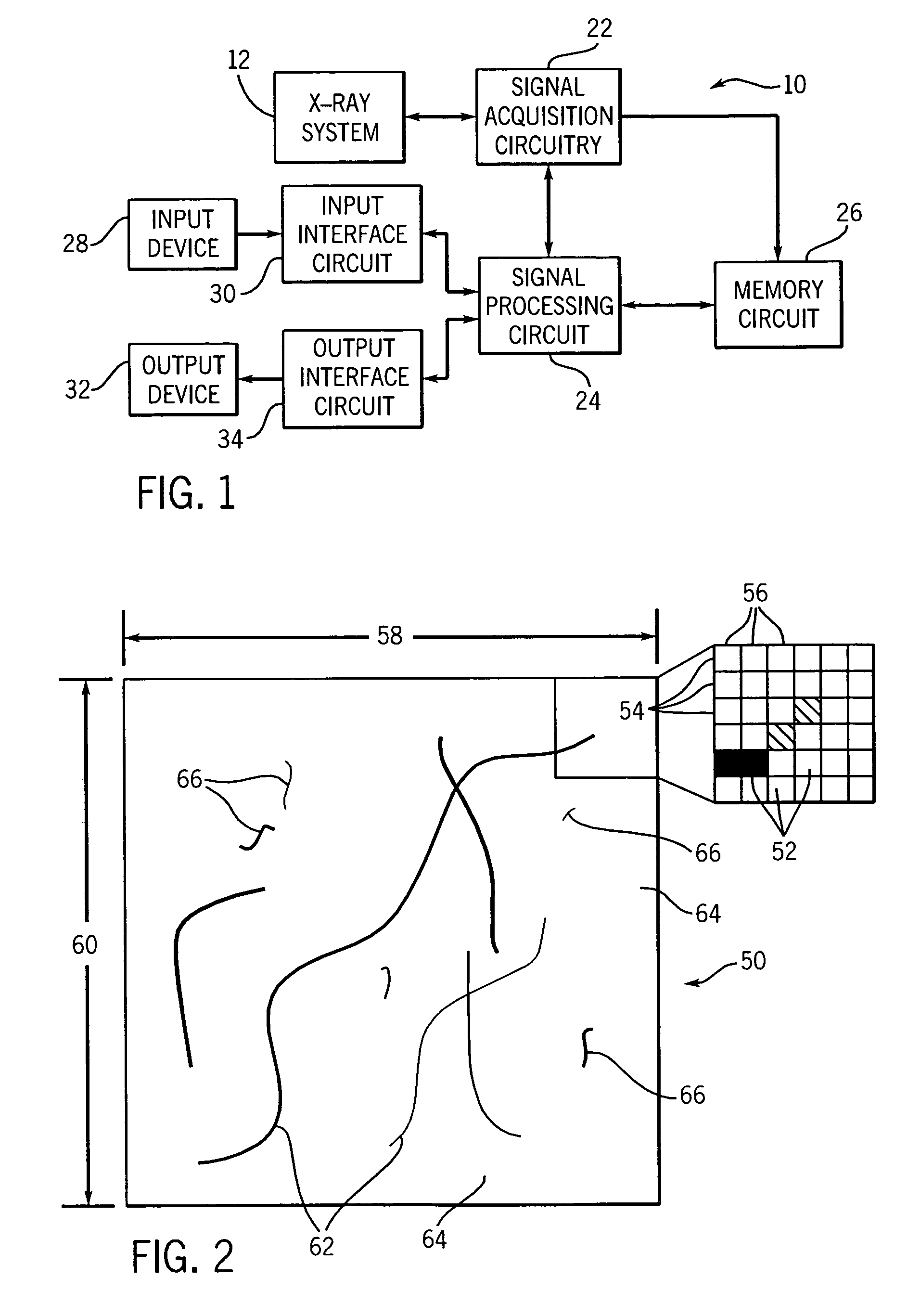
[0011]The invention provides an improved technique for enhancing discrete pixel images obtained from X-ray dual energy acquisition systems which is computationally efficient and which maintains image quality. The technique provides a means of combining multi-resolution decomposition (wavelet based processing) with segmentation based techniques which identify structures within an image and separately process the pixels associated with the structures. This combination allows the technique to exploit the redundancies of an image, as with wavelet based techniques, while also allowing the separate processing of structures and non-structures, as in segmentation-based techniques. Further, the technique provides a means of utilizing information in the high-energy image to assist in the identification and processing of structure in the both the bone image or the soft tissue image. In this way, information from both images may be utilized to improve the identification of structure within the bone image or soft tissue image. The combination of these techniques results in a computationally efficient, yet robust, noise reduction filter which may be applied to a variety of pixel based images.
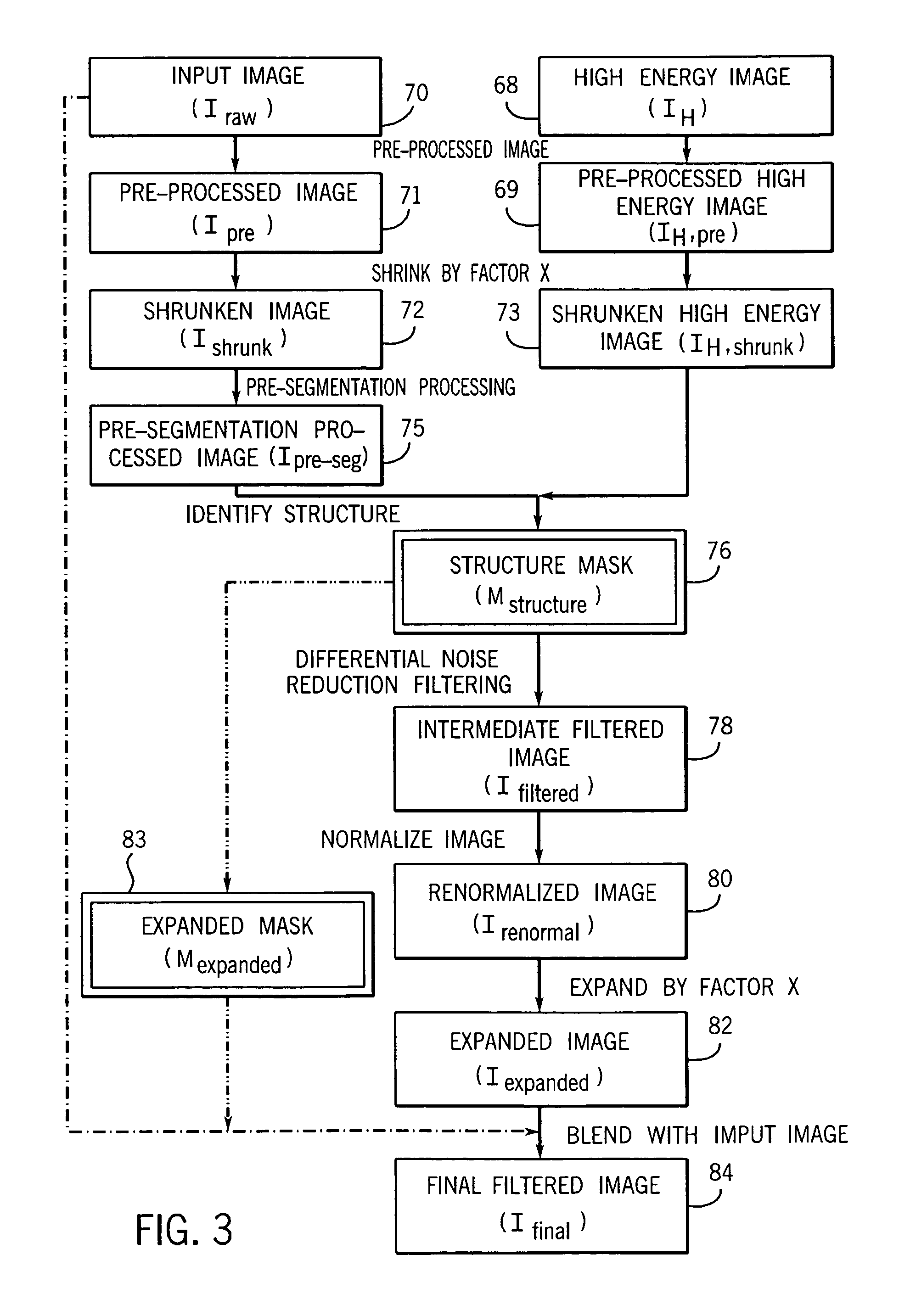
[0012]In an exemplary embodiment, multi-resolution decomposition occurs when a high-energy image and a bone image or a soft tissue image are shrunk by a given factor, allowing for the exploitation of redundancies in the images during subsequent processing. The shrunken bone image or soft tissue image is then processed using segmentation based techniques which begin by identifying the structure elements within a blurred or smoothed image. To further improve the identification of structure elements, complementary intensity information from the high-energy may also be used to determine the presence of structural pixels. Segmentation processing renders the structural details more robust and less susceptible to noise. A scalable threshold may serve as the basis for the identification of structural regions, making the enhancement framework inherently applicable to a range of image types and data characteristics. While small, isolated regions may be filtered out of the image, certain of these may be recuperated to maintain edge and feature continuity.
[0014]Upon completion of the segmentation based processing, the image is expanded by the same factor it was originally shrunk by to return it to its original size. Original texture may be added back to non-structural regions to further facilitate interpretation of both the non-structural and structural features. The ability of the present technique to increase computational efficiency, due to exploitation of the image redundancies, while maintaining image quality is particularly noteworthy since a reduction in image quality might be expected as a result of the image resizing. Surprisingly, however, no such decrease in image quality is observed.

Problems solved by technology

Typically one impediment to interpretation or further processing is the pixel to pixel variation which is attributable to acquisition noise.
While acquisition noise is usually random, there may also be additional structured noise as well which may be observed as artifacts in the image.
Moreover, while a number of X-ray image processing parameters may control the final image presentation, it is often difficult to determine which of these parameters, or which combination of the parameters, may be adjusted to provide the optimal image presentation.
Known signal processing systems for enhancing discrete pixel images suffer from certain drawbacks.
For example, such systems may not consistently provide comparable image presentations in which salient features or structures may be easily visualized.
Signal processing techniques employed in known systems are often difficult to reconfigure or adjust, owing to the relative inflexibility of hardware or firmware devices in which they are implemented or to the coding approach employed in software.
Moreover, current techniques may result in highlighting of small, isolated areas which are not important for interpretation and may be distracting to the viewer.
Conversely, in techniques enhancing images by feature structure recognition, breaks or discontinuities may be created between separate structural portions, such as along edges.
Such techniques may provide some degree of smoothing or edge enhancement, but may not provide satisfactory retention of textures at ends of edges or lines.
Finally, known signal processing techniques often employ inefficient computational noise reduction algorithms, resulting in delays in formulation of the reconstituted image or under-utilization of signal processing capabilities.

Method used


Image

  • Segmentation driven image noise reduction filter
  • Segmentation driven image noise reduction filter
  • Segmentation driven image noise reduction filter

Examples


Embodiment Construction

[0033]A highly abstracted rendition of image acquisition and processing by the preferred embodiment is illustrated in FIG. 17. FIG. 17 describes a preferred embodiment of the present technique whereby images are initially acquired by a dual energy X-ray system 12. The two acquired images are low energy image 67 and high energy image 68. Low energy image 67 is registered with respect to the high energy image in an optional motion correction process.

[0034]A dual energy subtraction process is then applied to registered low energy image 74 and to high energy image 68 to produce both a bone image 70 and a soft tissue image 70. Both the soft tissue image 70 and the bone image 70 are denoted by the same reference numeral because both images constitute an input image 70 which undergoes the claimed noise reduction process in the subsequent processes. Both input images 70 undergo noise reduction processing to yield final images 84, here denoted as final bone image 84 and final soft tissue ima...


PUM


Abstract

A technique for reducing noise in pixel images includes shrinking a first image and a second, related, image, and processing the first shrunken image with a novel segmentation-based filtering technique which utilizes image data from the second shrunken image to help identify structures within the image. After the structures are identified, the first shrunken image is differentially processed to reduce image noise. After processing, the first shrunken image is enlarged to the dimensions of the initial data, subsequently processed if necessary and the final image is displayed or analyzed. The resulting technique is versatile and provides greatly improved computational efficiency while maintaining image quality and robustness.

Description

FIELD OF THE INVENTION[0001]This invention relates to discrete picture element or pixel imaging techniques, and, more particularly, to an improved technique for analyzing and modifying values representative of pixels in X-ray images which significantly increases computational efficiency while maintaining overall image quality.BACKGROUND OF THE INVENTION[0002]A variety of discrete pixel imaging techniques are known and are presently in use. In general, such techniques rely on the collection or acquisition of data representative of each discrete pixel composing an image matrix. In the field of medical imaging, X-ray imaging techniques are available for producing the data represented by the pixels. In X-ray imaging, the pixel data is detected and encoded, such as in the form of digital values. The values are linked to particular relative locations of the pixels in the reconstructed image.[0003]The utility of a processed image is often largely dependent upon the degree to which it can b...

Claims


Application Information

Patent Timeline
IPC IPC(8): G06T5/00
CPCG06T5/002G06T5/50G06T7/0081G06T2207/10116G06T2207/20012G06T2207/20064G06T2207/20148G06T2207/30008G06T7/11G06T7/136G06T5/70
Inventor AVINASH, GOPAL B.
Owner GE MEDICAL SYST GLOBAL TECH CO LLC
Learn More