
Apparatus and method for stabilizing an implantable lead

a technology of implantable leads and apparatus, which is applied in the field of system and method for stabilizing the lead, can solve the problems of difficult removal of the lead, inconvenient use of tools, and inability to safely remove the lead, so as to promote tissue in-growth or attachment and enhance the stabilization effect of the implantable lead

Inactive Publication Date: 2005-11-01
CARDIAC PACEMAKERS INC
View PDF20 Cites 58 Cited by
  • Summary
  • Abstract
  • Description
  • Claims
  • Application Information

AI Technical Summary

Benefits of technology

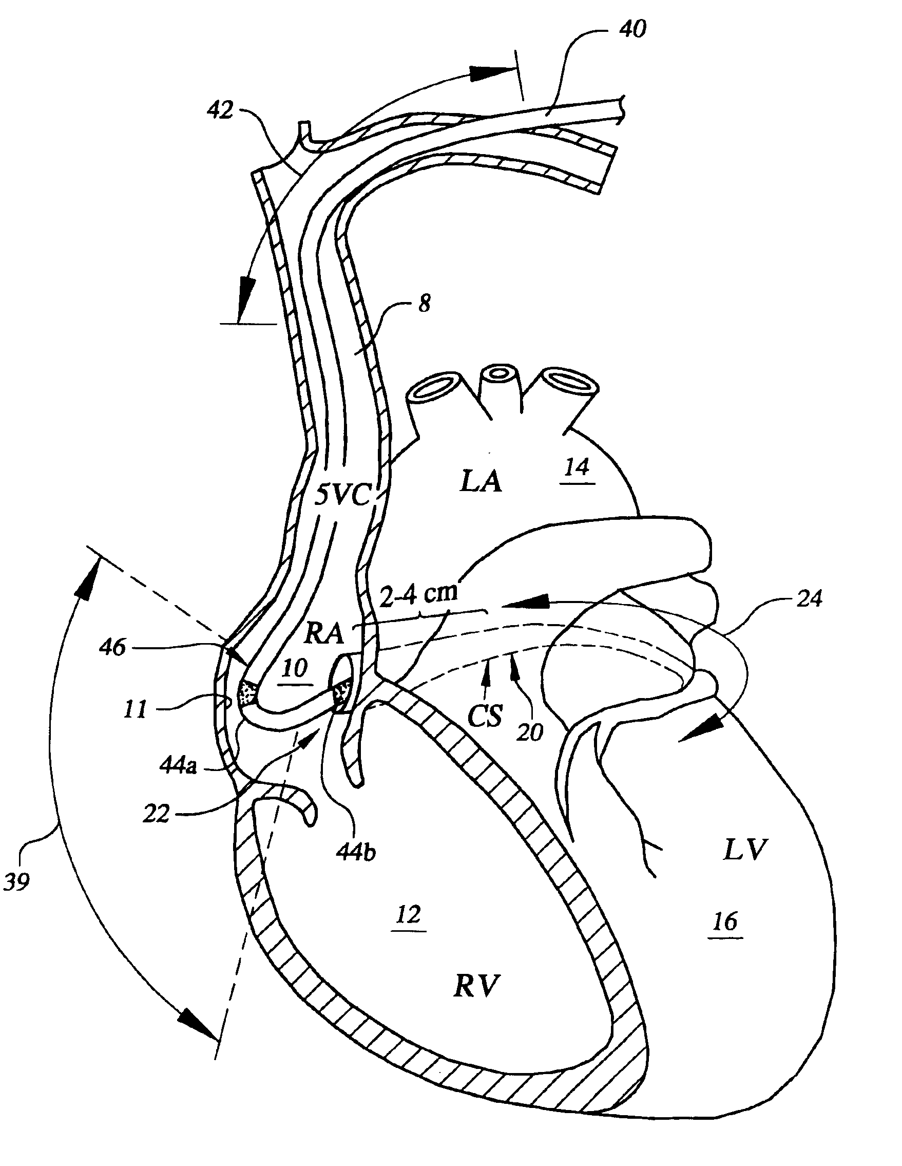
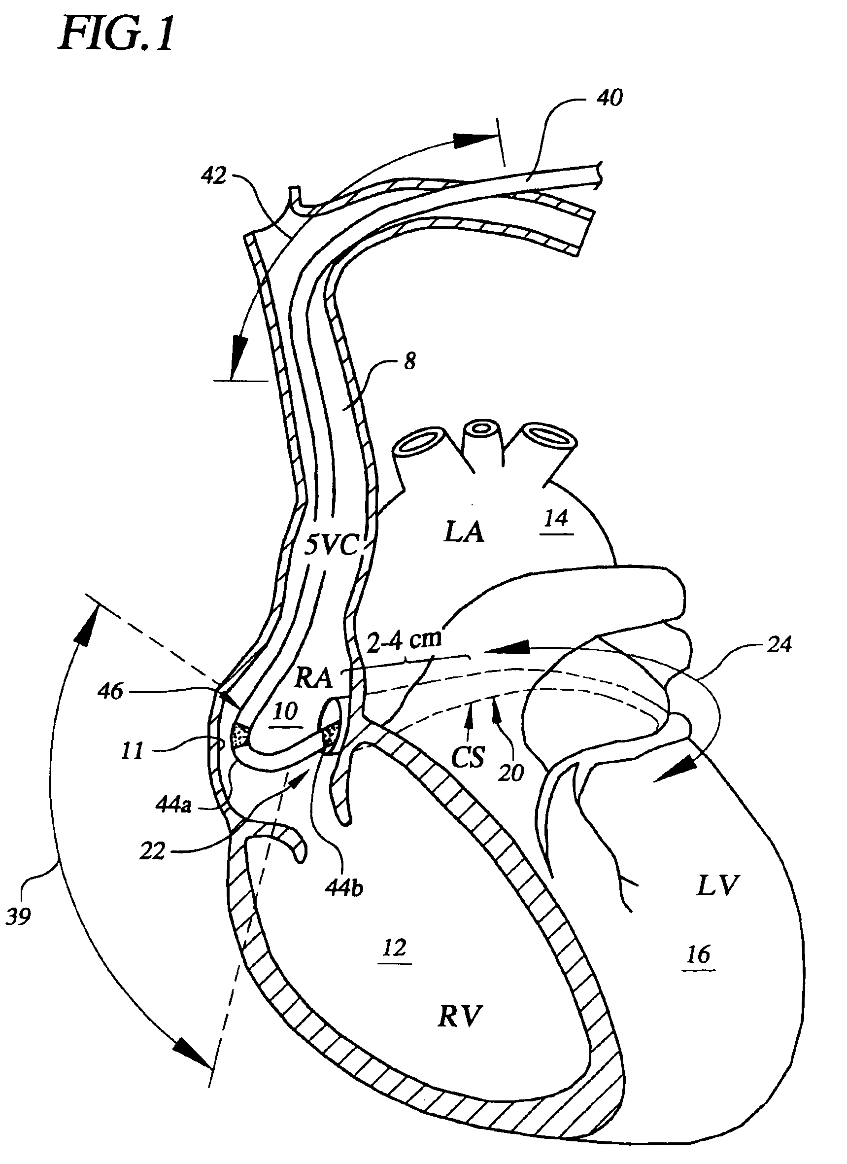
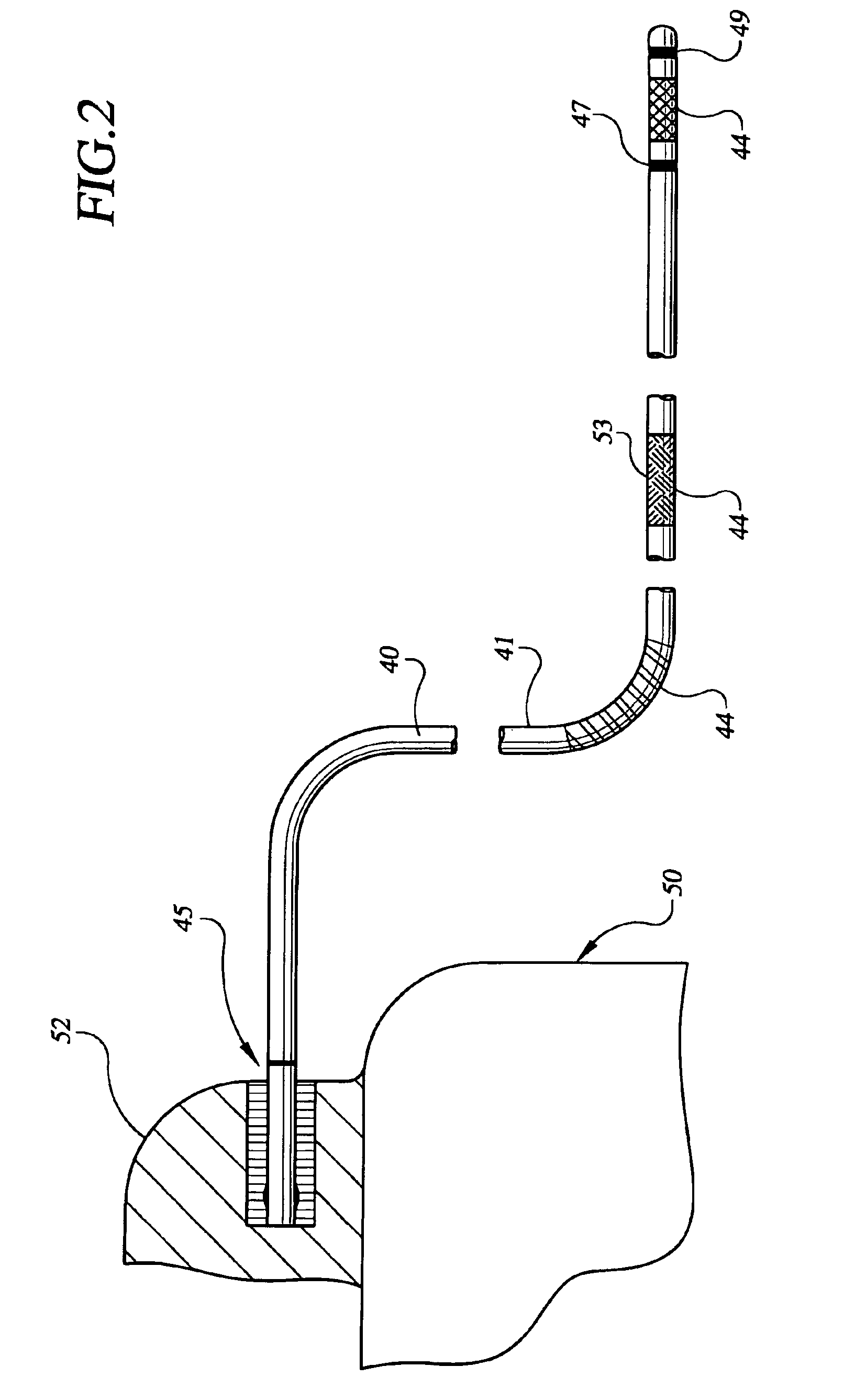
[0006]The present invention is directed to a system and method of stabilizing an implantable lead. The present invention provides particular advantages for leads designed to pass into or through the coronary sinus of a heart. According to one embodiment, the lead system includes a sleeve arrangement having one or more first locations comprising a first material that substantially prevents or inhibits tissue in-growth between the first locations and cardiac tissue contacting the first locations. The sleeve arrangement further includes one or more adhesion sites provided at one or more of the first locations. The adhesion sites promote tissue in-growth or attachment between the adhesion sites and cardiac tissue contacting the adhesion sites to enhance stabilization of the implantable lead. The cardiac tissue may represent tissue of a cardiac structure of the heart or coronary vasculature of the heart.
[0019]According to yet another embodiment of the present invention, a method of stabilizing a lead passing into a coronary sinus of a heart involves providing a sleeve arrangement on the lead. The sleeve arrangement includes one or more first locations comprising a first material and one or more adhesion sites provided at the one or more first locations. The method involves substantially preventing tissue in-growth between the first locations and cardiac tissue contacting the first locations. The method further involves promoting tissue in-growth or attachment between the adhesion sites and cardiac tissue contacting the adhesion sites to enhance stabilization of the lead passing into or through the coronary sinus. Electrical energy, such as pacing or defibrillating energy, may be produced at the adhesion sites. Electrical energy may also be sensed at the adhesion sites. Further, one or more physiologic parameters may be sensed at or proximate the adhesions sites.

Problems solved by technology

Removal of these leads is problematic, especially if coil electrodes are employed on the lead.
Presently, there are no extraction tools available for the safe removal of coronary vein leads.
Such tools, however, are not suited for use within thin walled vessels.
Presently available extraction tools, for example, can only be safely used to enter the proximal portions of the coronary sinus.
The risk of significant damage to the vasculature is very high, which can result in cardiac tamponade and death.
Consequently, physicians are presently limited to using locking stylets and simple traction as a means of removing coronary vein leads.
Although the extractability characteristics of leads can be improved using certain lead coatings, use of such coatings has been found to significantly reduce lead stability.
As such, the gains in lead extractability realizable through use of conventional lead coatings are achieved at the cost of reduced lead stability.

Method used


Image

  • Apparatus and method for stabilizing an implantable lead
  • Apparatus and method for stabilizing an implantable lead
  • Apparatus and method for stabilizing an implantable lead

Examples


Embodiment Construction

[0040]In the following description of the illustrated embodiments, references are made to the accompanying drawings which form a part hereof, and in which is shown by way of illustration, various embodiments in which the invention may be practiced. It is to be understood that other embodiments may be utilized, and structural and functional changes may be made without departing from the scope of the present invention.

[0041]In broad and general terms, the present invention is directed to a lead apparatus that provides for increased stability after implant and improved extractability when removal of the lead apparatus is needed or desired. One particular advantage of a lead apparatus implemented according to the principles of the present invention concerns the ability to control the stability and extractability characteristics provided by the lead apparatus. Another advantage concerns the provision of a primary lead fixation mechanism and a secondary lead fixation mechanism for stabili...


PUM


Abstract

A method and system for stabilizing an implantable lead system employs a lead having one or more sensing, pacing, or shocking electrodes. A sleeve arrangement of the lead includes one or more first locations comprising a first material that substantially prevents or inhibits tissue in-growth between the first locations and cardiac tissue contacting the first locations. The sleeve arrangement further includes one or more adhesion sites provided at one or more of the first locations. The adhesion sites promote tissue in-growth or attachment between the adhesion sites and cardiac tissue contacting the adhesion sites. The cardiac tissue may represent tissue of a cardiac structure of the heart or coronary vasculature of the heart.

Description

FIELD OF THE INVENTION[0001]The present invention relates generally to implantable leads and, more particularly, to systems and methods for stabilizing a lead in coronary sinus vasculature.BACKGROUND OF THE INVENTION[0002]Implantation of pacing and defibrillation leads in the coronary vessels is becoming increasingly common as atrial and heart failure therapies become more widely accepted. Implanting and stabilizing such leads in the coronary sinus, great vein, and the branch veins is critical to the efficacy of these and other therapies. It is often desirable or necessary to remove leads implanted in the coronary sinus vasculature for various reasons. Removal of these leads is problematic, especially if coil electrodes are employed on the lead.[0003]Presently, there are no extraction tools available for the safe removal of coronary vein leads. Various tools have been developed for removing right ventricular leads and right atrial leads, for example, such as mechanical dissection sh...

Claims


Application Information

Patent Timeline
Patent Type & Authority Patents(United States)
IPC IPC(8): A61N1/05
CPCA61N1/056A61N2001/0585
Inventor KRISHNAN, MOHANSCHEINER, AVRAMWESTLUND, RANDY
Owner CARDIAC PACEMAKERS INC
Learn More