
Instrument for regenerating living organism tissue or organ

a living organism and organ technology, applied in the direction of prosthesis, ligaments, blood vessels, etc., can solve the problems of inability to recover by grafting the cut nerve, loss of skin sensation, etc., of the area from ankle to instep, etc., and achieve the effect of efficient growth

Inactive Publication Date: 2005-10-11
UEDA MINORU
View PDF11 Cites 23 Cited by
  • Summary
  • Abstract
  • Description
  • Claims
  • Application Information

AI Technical Summary

Benefits of technology

[0011]An object of the present invention is to provide, in place of the conventional tubes for transplanting a nerve or a blood vessel, an instrument for regenerating a living organism tissue or organ which is free of the problem of a residual foreign matter in the body and the problem of a gap and into which the nerve, the blood vessel or the like can be readily inserted to enable cells to efficiently grow three-dimensionally.

Problems solved by technology

However, when the injured region is too large, restoration by the above-mentioned connection is impossible and it has been necessary to obtain a nerve from another location where a disorder, if any, could be believed to be less important than the disorder of the injured portion of concern and transplant it to the injured portion.
In this case, the problem is that usually skin sensation, etc. of the area from ankle to instep is lost.
In these experiments, although some cell proliferation is observed at the both ends of the cut nerve, no recovery by grafting the cut nerve is attained.
From this position they proliferate in such a direction that they cover the cut portion but mere covering of the cut portion leaves a gap between the cut ends and proliferation of cells is terminated before the cells completely fill the cut portion, so that restoration of the cut gap is not achieved.
Also, since the implanted tubular structure (also called a covering material) is artificially synthesized, it exists as a foreign matter in the body forever, which is undesirable.
Although use of a bioabsorbable material for the tubular structure solves the problem of residual foreign matter in the body, the problem of the existence of a gap still remains to be solved and it has been difficult to restore the deficit portion by cell proliferation.
That is, the collagen fiber bundle is thin and tends to be cut so that it is difficult to handle it and also it is difficult to insert it in a tubular structure such that it fully fills the inside of the tubular structure.
If the filling amount of the fiber bundle is decreased in order to facilitate insertion, there occur various defects that the gap between the fibers increases, the fiber bundle cannot be fixed well, localization of the fiber bundle inside the tubular member occurs and so on.
Therefore, both cases are undesirable since the gap inside is increased, giving the same results as those of the case where the above-mentioned gap is large.
Also, in the case where the filling amount of the fiber bundle is increased, other problems remain.
However, when the fiber bundle is inserted in a high filling density, exchange of substances is more inhibited closer to the central portion of the fiber bundle, so that it cannot be said that the environment is suitable for cell proliferation and, therefore, the fiber bundle is not suitable for restoring the nerve by cell proliferation.
However, when a growth factor or the like is coated on the surface of the lumen, the surface area is limited and the cell growth factor does not reach cells remote from the coated wall surface since the cells proliferate three-dimensionally.

Method used


Image

  • Instrument for regenerating living organism tissue or organ
  • Instrument for regenerating living organism tissue or organ
  • Instrument for regenerating living organism tissue or organ

Examples


example 1

Production of an Instrument for Regenerating a Living Organism Tissue or Organ

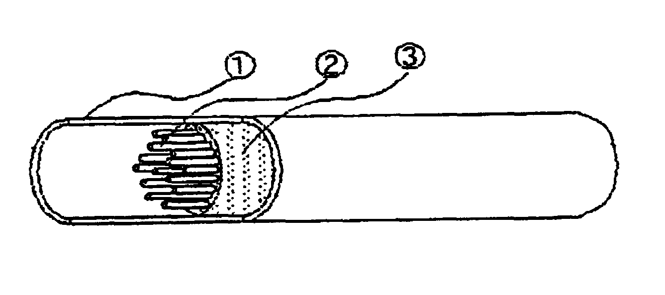
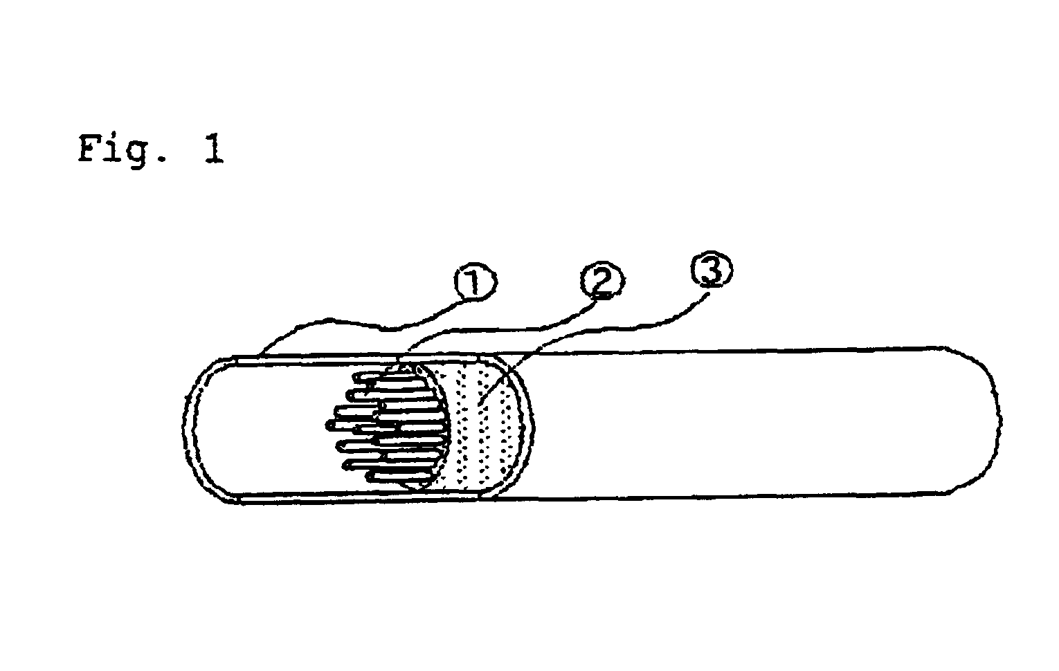
[0071]Enzyme-solubilized collagen (a mixture of collagen type I and type III) was dissolved in water to prepare an aqueous 5% solution and extruded in a coagulating liquid according to a conventional method to produce a collagen fiber having a diameter of 50 μm. The collagen fiber obtained was wound around a metal mandrel to produce a tubular structure constituted by the collagen and having an inner diameter of 1 mm and a thickness of 0.5 mm. In its lumen, 200 collagen fibers having a diameter of 50 μm were simultaneously inserted together with the aqueous 5% collagen solution and rapidly frozen and then lyophilization was performed in vacuo to produce a tubular instrument for regenerating a living organism tissue or organ entirely constituted by the collagen and of a structure such that it has a collagen fiber filling ratio of 50% in the lumen portion and a collagen-made porous material having a porosity ...

example 2

Tissue Regeneration Experiment on a Beagle

[0079]Using the regeneration instrument produced in Example 1, a tissue regeneration experiment on a beagle was conducted to confirm the regeneration of tissue under conditions close to that of humans. As for the tissue to be regenerated, a beagle peripheral nerve was selected.

[0080]The beagle fibular nerve was cut to make a 35 mm deficit portion. In this site the tubular collagen-made instrument for regenerating an organ mentioned above previously cut to 35 mm, i.e., the same length as the deficit portion length and subjected to 25-kGy γ-ray sterilization treatment was inserted and both ends thereof were sutured and fixed to the cut ends of the nerve with 8-0 polyamide based surgical suture at plural points.

Experimental Results

[0081]After the enthesis, an observation of nerve fiber regeneration was carried out by cutting out the tissue from the enthesis site at the time of the fifth week and performing an immunological tissue staining of pr...


PUM

PropertyMeasurementUnit
porosityaaaaaaaaaa
inner diameteraaaaaaaaaa

Abstract

This invention relates to an instrument for regenerating a living organism tissue or organ, characterized in that a support (A) formed from a biodegradable material or a bioabsorbable material includes a sponge-like fine matrix (B) formed from a biodegradable material or a bioabsorbable material and a linear guide channel (C) for a living organism tissue or organ.

Description

TECHNICAL FIELD OF THE INVENTION[0001]The present invention relates to an instrument for regenerating a living organism tissue / organ. More particularly the present invention relates to an instrument for regenerating a human tissue or organ, for example, a nerve fiber, a micro blood vessel, or the like, which was cut due to lesion or injury.BACKGROUND OF THE INVENTION[0002]In a case where a human tissue or organ such as a nerve or tendon is injured due to an accident, disaster or disease and the injury cannot be cured by self-recovery of a patient, a disorder occurs in perception, sensation, mobility or the like. For such a patient, with the development of a technology for connecting injured areas under a microscope in recent years, therapeutics such as surgical suturing for connecting cut portions or nerve autotransplantation, by which a nerve or tendon of a patient himself or herself biopsied from another part of the body is transplanted to recover the lost function, has been effec...

Claims


Application Information

Patent Timeline
Patent Type & Authority Patents(United States)
IPC IPC(8): A61F2/02A61F2/04A61F2/06A61F2/08A61L27/48A61L31/12
CPCA61L27/48A61L31/129C08L89/06
Inventor DOI, NOBUTOSHIMATSUDA, KAZUHISAHATA, KEN-ICHIROSAKAI, KENSUKE
Owner UEDA MINORU
Learn More