
System and method for directing and monitoring radiation

a radiation and radiation monitoring technology, applied in the field of radiation monitoring system and method, can solve the problems of not always being able to predict the dose of the actual entrance and exit location, the effect of radiation on normal cells cannot be overlooked, and the dose applied to the treated location, where interpolation has been performed, cannot always be predicted from the measured entrance and exit dos

Inactive Publication Date: 2002-08-13
REMON MEDICAL TECH
View PDF46 Cites 239 Cited by
  • Summary
  • Abstract
  • Description
  • Claims
  • Application Information

AI Technical Summary

Problems solved by technology

However, the effect of radiation on normal cells cannot be overlooked.
However, measurement of entrance and exit doses combined with interpolation can only predict the dosage of the actual entrance and exit locations which are directly measured.
In addition, the dosage applied to the treated locations, where interpolation has been performed, cannot always be predicted from the measured entrance and exit doses.
Unfortunately, such devices are not designed to relay data outside the patient's body.
However, this procedure provides the treating physician with very little useful information on the actual dosage delivered deep into the affected tissue and the surrounding body tissues.
Although such a system can incorporate many TLD devices and as such, achieve many measurements, the positioning of the adapter, and the size and shape of the adapter is not necessarily repeatable and does not necessarily correspond to the actual positioning, size and shape of the patient's breast and surrounding tissue.
In any case, radiation phantoms are localized outside the patient's body, and as such provide little information, if any, relating to the actual doses applied to the treated site.
Since most of the above mentioned monitoring methods rely heavily on mathematical calculations and projections, such methods fail to accurately predict the doses absorbed by various body regions.
Furthermore, since most of these methods employ calculations effected on information retrieved from the dosimetric points, field distortions, which can occur when a radiation beam or beams pass through an organ or tissue are not accounted for.
As such when monitoring is not effected directly in the tumor but rather on the skin, or on an extracorporeal phantom such as with the methods described above, the accurate prediction of dosage to the tumor itself ad to surrounding healthy tissues is not possible.
This method has very poor resolution, does not yield the dose locally absorbed by any specified organ and does not enable a closed loop control of the radiation conditions.
In addition, while using microwave radiation a secondary effect caused by the tissue heating resultant from the procedure is a structural modification of the tissue which can lead to a change of the path previously set for the therapeutic radiation.
Although x-ray fluoroscopy is used in combination with these procedures to direct the physician to the site of treatment, this method is limited to stationary treatment sites and as such the application of a gamma knife or a highly focused microwave beam to a treatment site of moving organs such as a pulsating heart cannot be easily effected with accuracy.
Such a device is, therefore, usable only for a very short time period and has a very short shelf life before use.
Moreover, the implantation of a radioactive stent or graft requires both an oncologist and a cardiologist to be present at the time of the procedure further complicating matters.
Thus, there exists no efficient system or method with which a physician can precisely direct a radiation dose of any radiation type to a treatment site and / or monitor with accuracy the radiative dosages absorbed by both the treatment site and the tissue which surrounds the treatment site.
First, if designed for prolonged implantation such wiring is prone to cause infections.
Second, such wiring is complicated to implement especially in cases where sensing of radiation from deep within the body is required.
It will be appreciated that implantable power source dependent telemetry has limitations inherent to the use of an exhaustible power source.
In addition, power sources are space consuming which is less favorable when miniaturization is advantageous, such as in the case of intrabody implanted sensors.
It is well known that the signal generatable by an implantable radio frequency transducer is rather weak, necessitating the implantation thereof close to the skin.
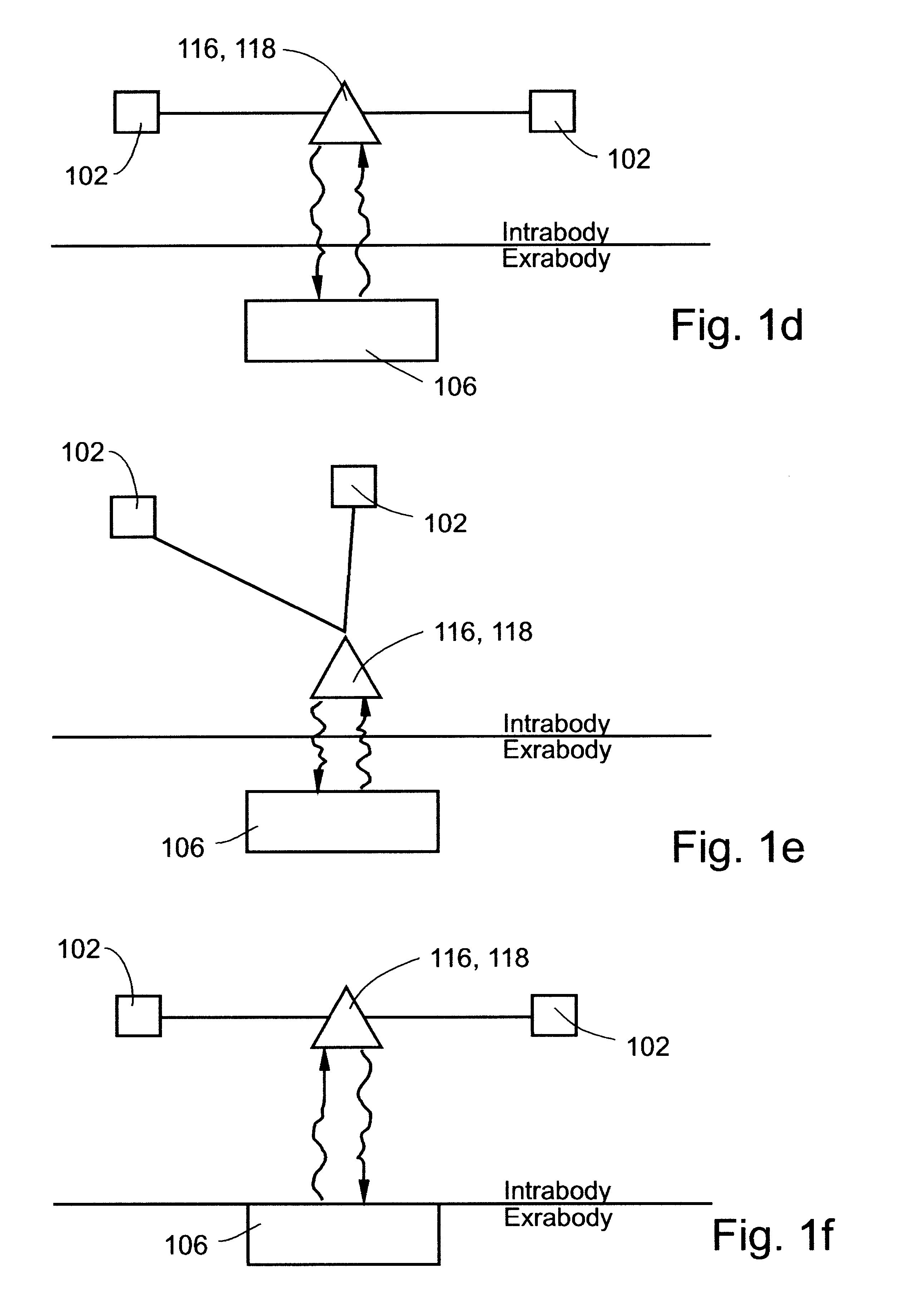
This however is limitative in case where radiation sensing is required deep within the body because complicated (long) wiring, as shown in FIG. 1e, is then required.
In addition, when using radio frequency telemetry, antenna(s) for receiving / transmitting radio frequency signals should be employed which is cumbersome, space consuming and highly directional dependent.
In this case, monitoring the ultrasonic radiation is only provided for the tissues surrounding the stones such as tissues of the kidney or gall bladder since implantation of system 100 within such stones is neither feasible nor is it necessary.

Method used


Image

  • System and method for directing and monitoring radiation
  • System and method for directing and monitoring radiation
  • System and method for directing and monitoring radiation

Examples


Embodiment Construction

Reference is now made to the following example, which together with the above descriptions, illustrate the invention in a non limiting fashion.

For purposes of better understanding the construction and operation of an acoustic transducer utilizable by the system of the present invention, reference is made to the construction and operation of an acoustic transducer as described in U.S. patent application Ser. No. 09 / 000,553.

Referring again to the drawings, FIGS. 5a, 5b and 6a-6e illustrate a preferred embodiment of a transducer element according to the present invention. As shown in the Figures, a transducer element 1 includes at least one cell member 3 including a cavity 4 etched into a substrate and covered by a substantially flexible piezoelectric layer 2. Attached to piezoelectric layer 2 are an upper electrode 8 and a lower electrode 6, the electrodes for connection to an electronic circuit.

The substrate is preferably made of an electrical conducting layer 11 disposed on an elect...


PUM


Abstract

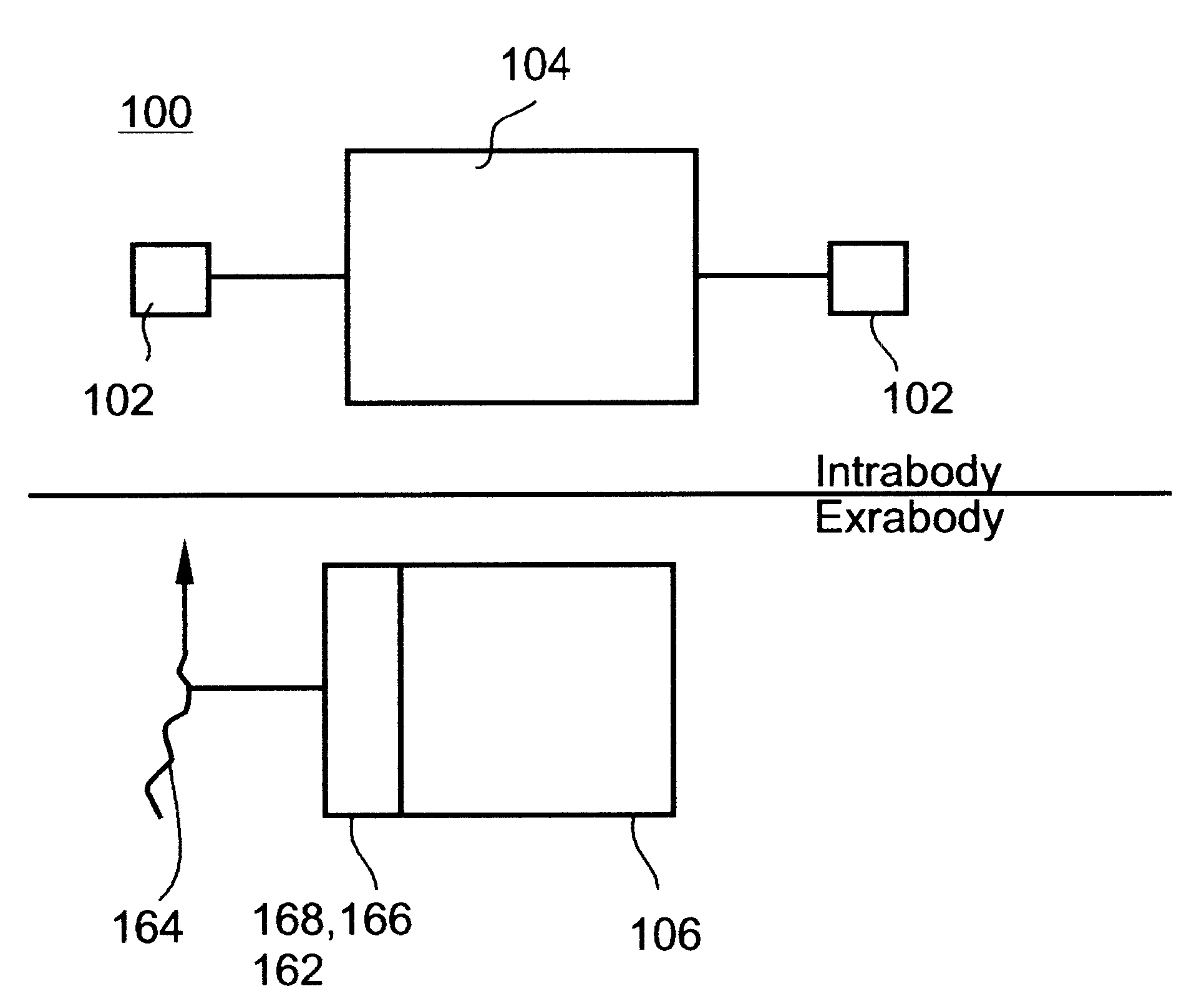
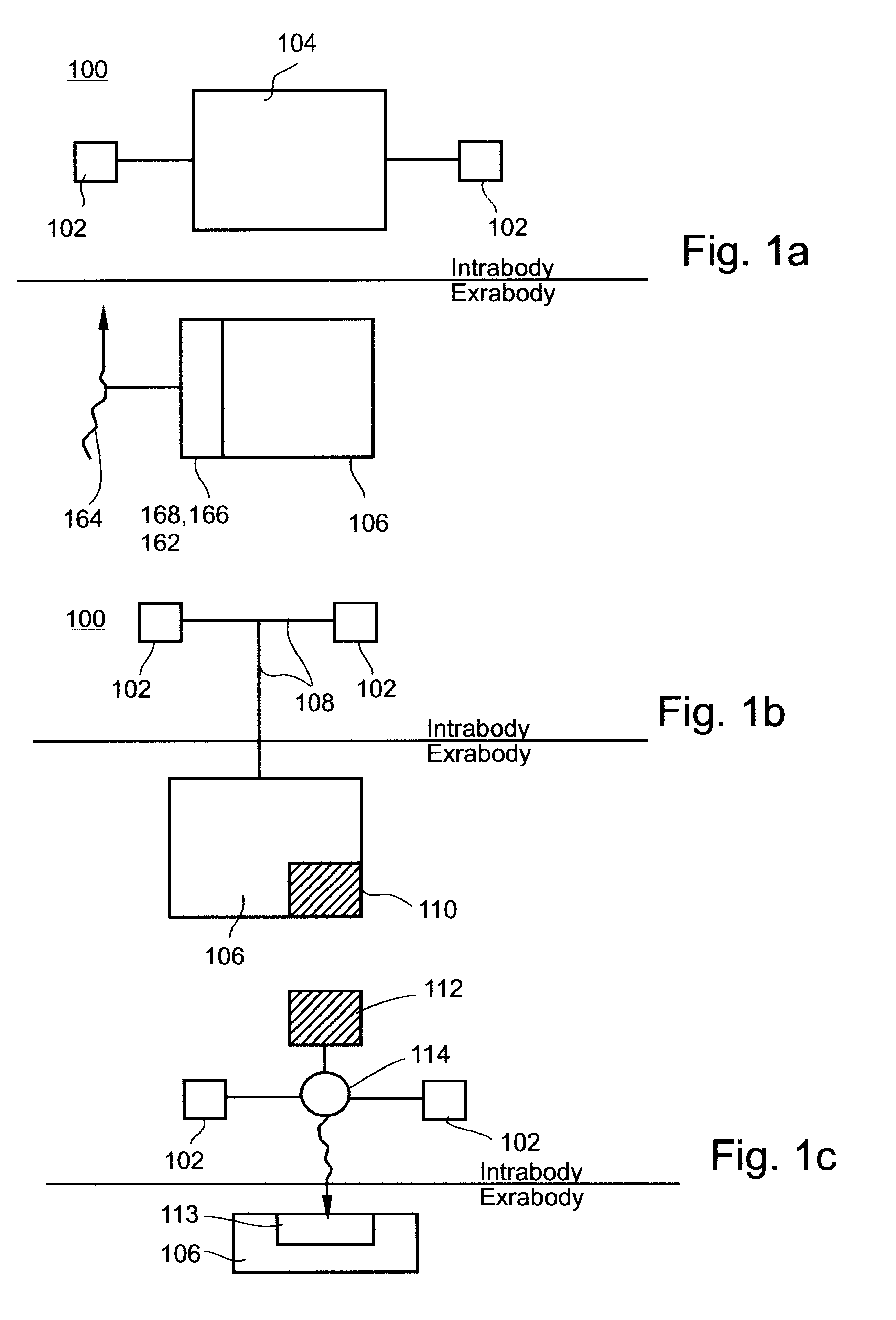
A system for monitoring, directing and controlling the dose of radiation in a medical procedure for irradiating a specific region of a patient's body. In its generic form, the system includes at least one sensor being implantable within, or in proximity to, the specific region of the patient's body, the at least one sensor being for sensing at least one parameter associated with the radiation. The system further includes a relaying device which is in communication with the sensor(s). The relaying device serves for relaying the information outside of the patient's body.

Description

FIELD AND BACKGROUND OF THE INVENTIONThe present invention relates to a system and method for intrabody directing and monitoring doses of therapeutic radiation applied to a patient's body. More particularly, the system and method of the present invention employs an implantable sensor coupled to a relaying device, with which, information pertaining to radiation sensed and optionally quantified by the sensors can be relayed outside the body. In addition, the system and method of the present invention can be utilized to assist in directing radiation to a treatment site within the patient's body.Radiation therapy is used extensively in the medical field to treat a variety of medical conditions. Radiation therapy typically utilizes electromagnetic radiation, typically ionizing radiation, such as, but not limited to, x-ray, gamma-rays and particle beams, as well as ultrasonic radiation to treat many types of cancers, tumors and, cell proliferative disorders as well as non-malignant medica...

Claims


Application Information

Patent Timeline
Patent Type & Authority Patents(United States)
IPC IPC(8): A61B5/053A61B5/00G01F1/34G01F1/72G01F1/36H01L41/113H04R17/00G01T1/16A61B17/22A61B18/00A61B19/00A61F7/00A61N5/02A61N5/06A61N5/10G01T1/161G01T1/17H03H9/17H10N30/00H10N30/20H10N30/30
CPCA61N5/1048H01L41/1132A61B5/0031A61B5/413A61B2562/028A61N1/37288A61N5/1071A61N2005/1072H04R17/00H10N30/302
Inventor PENNER, AVIPORAT, YARIVDORON, EYAL
Owner REMON MEDICAL TECH
Learn More