
Large area array single exposure digital mammography

a digital mammography and large-area array technology, applied in the field of digital radiology, can solve the problems of poor contrast of film mammograms, single traditional film x-ray exposure often does not show the entire tonal range of breast tissue of patients, and the inability to provide adequate high-quality images to detect cancer

Inactive Publication Date: 2002-07-09
TREX MEDICAL CORP
View PDF20 Cites 9 Cited by
  • Summary
  • Abstract
  • Description
  • Claims
  • Application Information

AI Technical Summary

Problems solved by technology

However, these techniques do not always provide adequately high-quality images to detect cancer, particularly in the relatively large population of women having radiodense breast tissue (younger women, for example, tend to have radiodense breast tissue).
As a result, a single traditional film x-ray exposure often does not show the entire tonal range of a patient's breast tissue.
Other drawbacks are caused by the poor contrast of film mammograms.
One of these drawbacks is that it is difficult to detect masses in patients having breast implants.
A second drawback is that it is difficult to discern between benign and malignant microcalcifications and tumors.
This latter drawback results in thousands of unnecessary invasive procedures to remove growths which are later determined to be benign.
One reason for this is because the electronic imagers for capturing the image are small and cannot be arranged together to provide a gap free imaging surface large enough to image an entire breast in a single exposure.
This one square inch area is not sufficient to image an entire breast.
Digital mammography also faces severe technical challenges.
Because of the large number of pixels needed to image an entire breast and electronic detector size limitation described above, these mammography machines use one of two systems to image an entire breast.
The scan time is long, which is uncomfortable for the patient and increases the likelihood of image discontinuity due to patient movement during the procedure (including involuntary motions such as breathing and heartbeats), adversely affecting the image quality.
The patient may be exposed to radiation longer than necessary for imaging, which is undesirable.
The mechanical devices necessary to maintain the beam / CCD array synchronization is complex and requires a special support system.
Therefore, this device cannot be used interchangeably with standard film / screen mammography machines.
Also, the x-ray tube is on for a prolonged time period, and is subjected to high heat loading.
Tungsten targets operate at suboptimal high kV ranges (near 40 kVp), which detrimentally affects the contrast between adipose (fatty), glandular, or calcified breast tissue.
As noted above, a low image contrast is unsuitable for imaging soft tissue such as breasts.
The mechanics of this device preclude it from being used interchangeably with standard film / screen mammography machines.
This method also has the disadvantage that the result is several images, not a single image, of the breast.

Method used


Image

  • Large area array single exposure digital mammography
  • Large area array single exposure digital mammography
  • Large area array single exposure digital mammography

Examples


Embodiment Construction

1. A Single Exposure, Full Field Digital Array

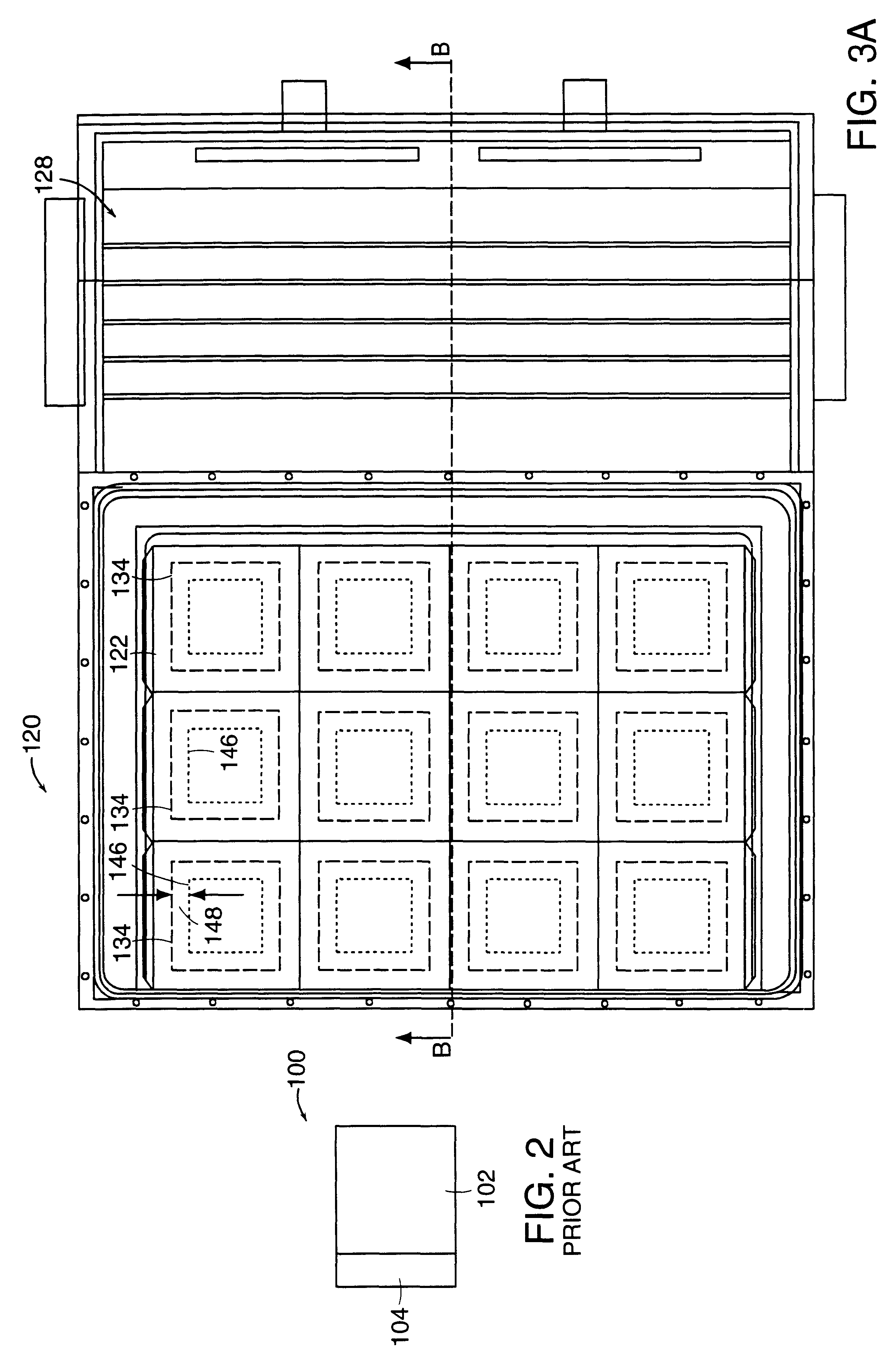
FIGS. 3A and 3B illustrate a preferred embodiment of a single exposure, large area imager array 120. The imager array is possible by a novel technique for butting together a number of electronic imagers, such as those shown in FIG. 2, without a gap in the image. The preferred embodiment employs CCD (charged coupled device) sensors, but a person skilled in the art understands that other electronic imagers, such as amorphous polysilicon flat panel detectors, selenium storage plates, zinc cadmium telluride sensors, and the like may be used as well.

The single exposure, large area imager array 120 provides an image receptor surface 122 having no significant gaps. The imager array comprises an image receptor 124, a thermoelectric cooling arrangement 126, and receptor electronics 128. The image receptor 124 comprises a scintillator 130, an optical system 132, such as a fiber optic bundle, and an electronic imager 134, such as CCDs. The thermoel...


PUM


Abstract

A unique digital imaging device includes the digital imager, a mammography machine including the digital imager, a method for processing the digital image obtained by the digital imager, a local area network (LAN) comprising a number of mammography machines and one or more work stations, and a wide area network (WAN) for transmitting digital images to remote locations. The digital imager according to the present invention comprises a large area, single exposure digital imaging device is provided that allows a number of electronic imagers to be butted together to form an array of electronic imagers large enough to image an entire breast in a single exposure. A preferred embodiment of the digital imager comprises an optical system, such as fiber optic bundles, optically coupled with the surface of the electronic imager. The optical systems may have a surface larger than the electronic sensors, and may be butted together to provide a gap-free imaging surface.

Description

1. Field of the InventionThe present invention relates to digital radiology and, more particularly, to a large array, single exposure digital mammography machine. The digital mammography machine may process, store access, and transmit data in the same manner as other types of digital image data.2. Discussion of the Prior ArtAccording to the American Cancer Society, in 1995 182,000 women will be diagnosed with breast cancer and 46,000 women will die from breast cancer. The ACS estimates that 2,000,000 women will be diagnosed and more than 500,000 women will die of breast cancer in the 1990's. Early breast cancer detection increases the patient's chances of surviving the cancer. Thus, early detection is a major factor in saving the lives of breast cancer patients. Experts agree that a mammogram is the single best means of early breast cancer detection.Mammography is the radiological examination of the human breast. It is generally accepted that mammography is an effective and reliable...

Claims


Application Information

Patent Timeline
IPC IPC(8): A61B5/00A61B6/00
CPCA61B6/4233A61B6/4488A61B6/502A61B6/548A61B5/002
Inventor CHEUNG, LIM
Owner TREX MEDICAL CORP
Learn More