
Genetically encoded fluorescent indicators under optogenetic control and uses thereof

a fluorescent indicator and optogenetic control technology, applied in the field of artifact-free alloptical characterization of cellular physiology, can solve the problems of limiting the use of red gecis in conjunction with blue-light activated optogenetic actuators, reducing the sensitivity of rcamp variants, and complicated photophysics and lysosomal accumulation of r-geco variants

Pending Publication Date: 2022-01-20
LUMISTAR BIOTECHNOLOGY INC +1
View PDF0 Cites 0 Cited by
  • Summary
  • Abstract
  • Description
  • Claims
  • Application Information

AI Technical Summary

Benefits of technology

The present invention is related to the use of genetically encoded Ca2+ indicators (GECIs) for studying calcium dynamics in cells. The invention includes methods for drug efficacy and toxicity assessment, as well as for toxicological screening and monitoring of cell functions under different conditions. The invention uses red-shifted GECIs that are activated by light having different wavelengths than the ones used for stimulation. The method involves expressing the red-shifted GECI in the cell and obtaining an optical signal in response to stimulation. Additionally, the invention includes using optical actuators that are activated by light in a different range than the GECI, and co-expressing them with the GECI in the cell. This allows for the study of cell functions with improved specificity and accuracy.

Problems solved by technology

However, as widely recognized, red GECIs currently suffer from a number of limitations compared to the most highly optimized green GECIs (i.e., GCaMP6).
These limitations include decreased sensitivity for RCaMP variants and complicated photophysics and lysosomal accumulation for R-GECO variants, especially the inherited property of undesirable blue-light activated photoswitching behavior that was also present in the DsRed-derived template (mApple) from which they were engineered.
Such an artifact limits their use in conjunction with blue-light activated optogenetic actuators for all-optical stimulation and observation.
While existing indicators with longer excitation and emission wavelengths falling within the near infrared (NIR) optical window (˜650 nm to 900 nm) are ideal for in vivo imaging due to the minimum tissue scattering and absorption, the critical problem they have encountered is the dim fluorescence for qualified imaging acquisition.

Method used


Image

  • Genetically encoded fluorescent indicators under optogenetic control and uses thereof
  • Genetically encoded fluorescent indicators under optogenetic control and uses thereof
  • Genetically encoded fluorescent indicators under optogenetic control and uses thereof

Examples


example 1

for Mammalian Cell Imaging and Virus Production

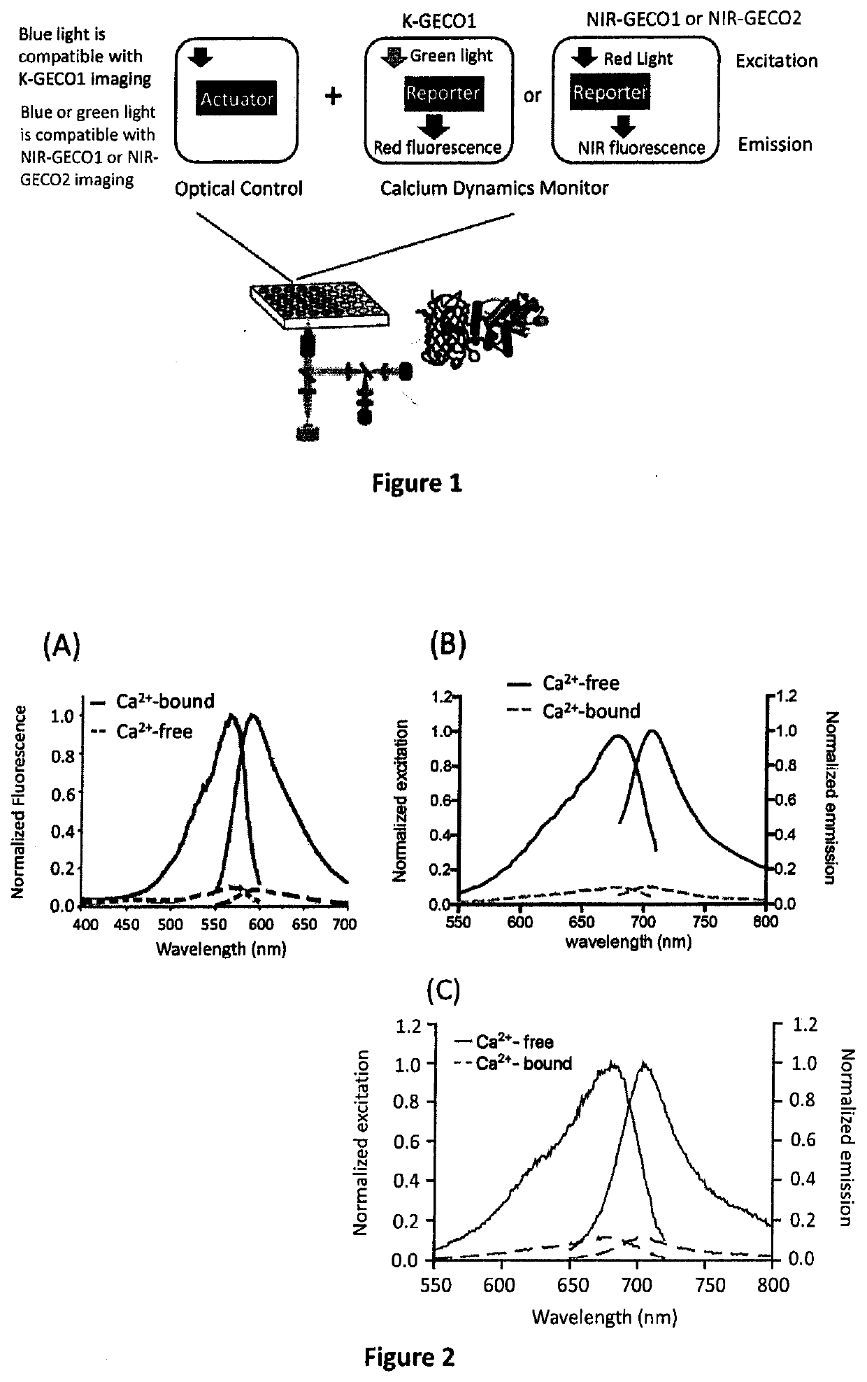
[0060]ChR2-EYFP linked by a 2A peptide to K-GECO1 was amplified and subcloned into the pshuttle backbone for commercial adenoviral production (Vector Biolabs, Malvern, Pa., US). All sequences are provided in Example 7.

example 2

Characterization

[0061]To purify K-GECO variants for in vitro characterization, the pBAD / His B plasmid encoding the variant of interest was used to transform electrocompetent E. coli DH10B cells and then plated on LB-agar plate with ampicillin (400 μg / mL). Single colonies were picked and inoculated into 5 mL LB medium supplemented with 100 g / mL ampicillin. Bacterial subcultures were incubated overnight at 37° C. Then, 5 mL of bacterial subculture was added into 500 mL of LB medium with 100 μg / mL of ampicillin. The cultures were incubated at 37° C. to an OD of 0.6. Following induction with L-arabinose to a final concentration of 0.02% (wt / vol), the cultures were then incubated at 20° C. overnight. Bacteria were harvested by centrifugation at 4000 g for 10 min, resuspended in 30 mM Tris-HCl buffer (pH 7.4), lysed using a French press, and then clarified by centrifugation at 13,000 g for 30 mins. Proteins were purified from the cell-free extract by Ni-NTA affinity chromatography (MCLAB)...

example 3a

re Conditions of HL-1 Cell Line

[0067]To culture the HL-1 cell line, flasks were pre-coated with gelatin / fibronectin at 37° C. overnight. Cells were cultured in supplemented Claycomb Medium (Claycomb Medium with 10% fetal bovine serum (Sigma Aldrich 12103C (Batch 8A0177)), 1 U / ml penicillin / streptomycin, 0.1 mM norepinephrine and 2 mM L-glutamine) and split 1:3 when they reached confluency.


PUM

PropertyMeasurementUnit
excitation wavelengthaaaaaaaaaa
emission wavelengthsaaaaaaaaaa
activation wavelengthaaaaaaaaaa

Abstract

Disclosed are methods and compositions for real-time monitoring cellular calcium ion dynamics by red-shifted genetically encoded indicators under optogenetic control, in which millisecond-timescale of temporal control of optical activation or inactivation and signal recording can be achieved. The methods include artifact-free functional imaging in conjunction with optogenetic tools for studying cellular physiology, signal transduction and neuronal activity. Thus, all-optical and non-invasive approaches for drug screening, toxicity testing and assessment of cell functions may be provided.

Description

FIELD OF THE INVENTION[0001]The present invention relates to methods and compositions related to artifact-free all-optical characterization of cellular physiology, which may be used for drug screening, toxicity testing and assessment of cell functions.BACKGROUND[0002]The calcium ion (Ca2+) is a versatile second messenger, playing an essential role in all aspects of physiology and controlling key cell fate decisions, especially in excitable cells, such as cardiomyocytes, neurons and so on. Thus, the kinetics and spatial properties of Ca2+ signals in cellular or subcellular compartments can reveal physiological response of cells to external stimuli for interrogation.[0003]The use of genetically encoded Ca2+ indicators (GECIs) has proven to be an indispensable tool for studying the spatio-temporal dynamics of cellular Ca2+ signals in vivo and in vitro. They have been adopted as chronic probes for simultaneously monitoring neuronal activity in contexts ranging from dissociated neurons i...

Claims


Application Information

Patent Timeline
Patent Type & Authority Applications(United States)
IPC IPC(8): C12Q1/6897G01N21/64
CPCC12Q1/6897G01N21/6428C07K14/415
Inventor CHANG, YU-FENSHEN, YIQIAN, YONGCHUNG, MIN-WENCAMPBELL
Owner LUMISTAR BIOTECHNOLOGY INC
Learn More