
Eye Phantom for Evaluating Retinal Angiography Image

a retinal angiography and eye phantom technology, applied in the field of eye phantom for evaluating retinal angiography images, can solve the problems of difficult systematic evaluation inability to accurately evaluate the performance and inability to perform the process of checking and improving performance in the development process of domestic oct equipment. , to achieve the effect of accurately evaluating the performance of the equipment under development and evaluating the performan

Pending Publication Date: 2022-01-06
KOREA RES INST OF STANDARDS & SCI
View PDF0 Cites 0 Cited by
  • Summary
  • Abstract
  • Description
  • Claims
  • Application Information

AI Technical Summary

Benefits of technology

The present invention is about an eye phantom that includes fine fluid channel structures that mimic the vascular structure of the actual retina. This makes the eye phantom more accurate and useful for evaluating the performance of ophthalmic equipment. The eye phantom can also be used to evaluate the performance of a fluorescent blood vessel contrast fundus camera. The technical effect is that the eye phantom provides a reliable and accurate method for evaluating the performance of ophthalmic equipment.

Problems solved by technology

However, Korea currently imports almost all of the equipment from overseas companies such as the United States, Germany and Japan, and it has been pointed out that the domestic OCT technology field is too dependent on foreign technology.
However, there are the following problems in developing domestic OCT equipment.
That is, there is a problem in that it is still difficult to perform the systematic evaluation on the OCT equipment in Korea, and the process of checking and improving performance in the process of developing domestic OCT equipment is not smoothly performed.
However, since it is impossible to conduct tests on animals or humans in the process of product development or in the process of performance evaluation and production for obtaining medical device certification, it is very difficult to realize performance verification and improvement work in the equipment development process.
Therefore, it is impossible to obtain an image corresponding to an angiography image from an OCT image of a conventional eye phantom, and there is a problem in that there is a limitation in applying the conventional eye phantom to the development of the fifth generation OCT.

Method used


Image

  • Eye Phantom for Evaluating Retinal Angiography Image
  • Eye Phantom for Evaluating Retinal Angiography Image
  • Eye Phantom for Evaluating Retinal Angiography Image

Examples


Embodiment Construction

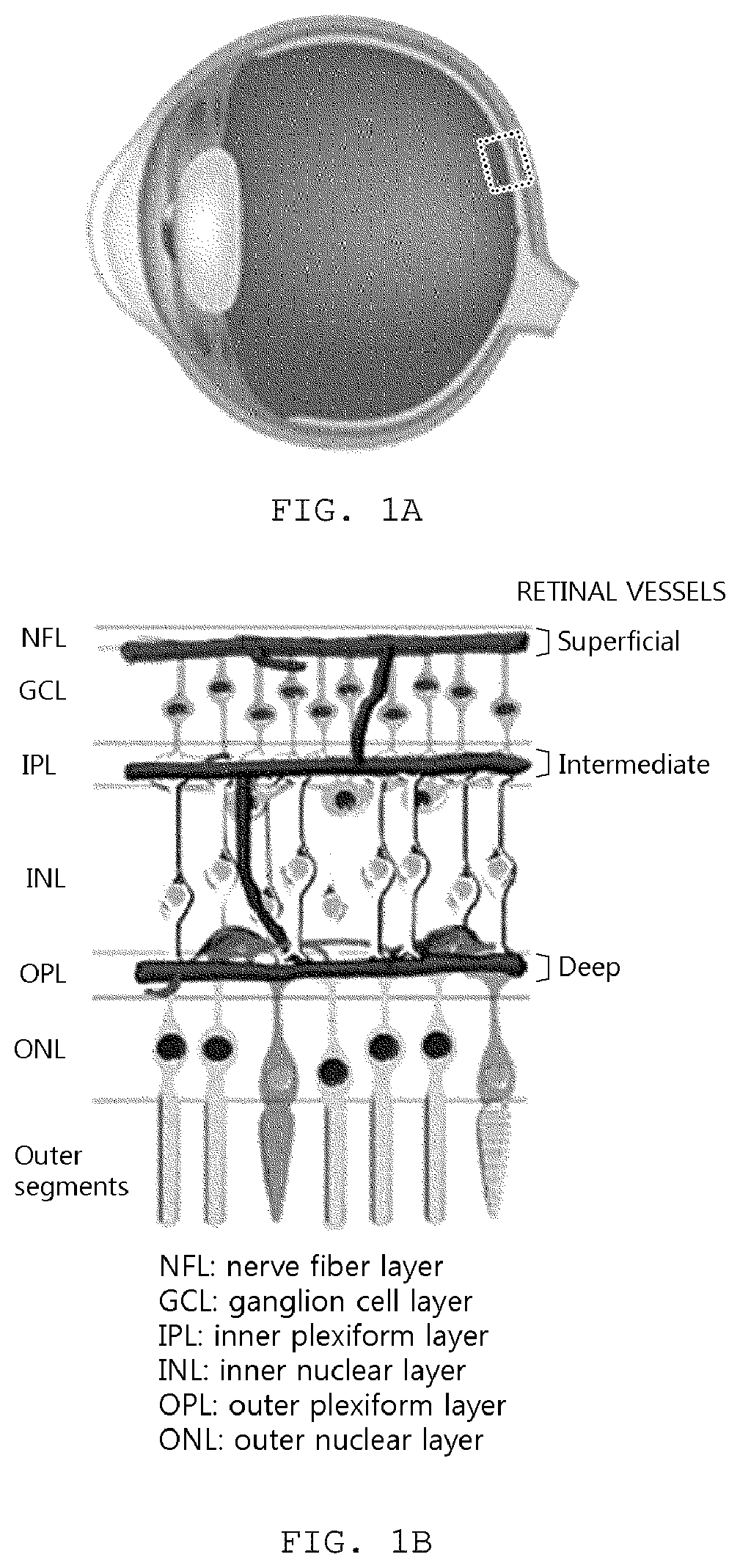
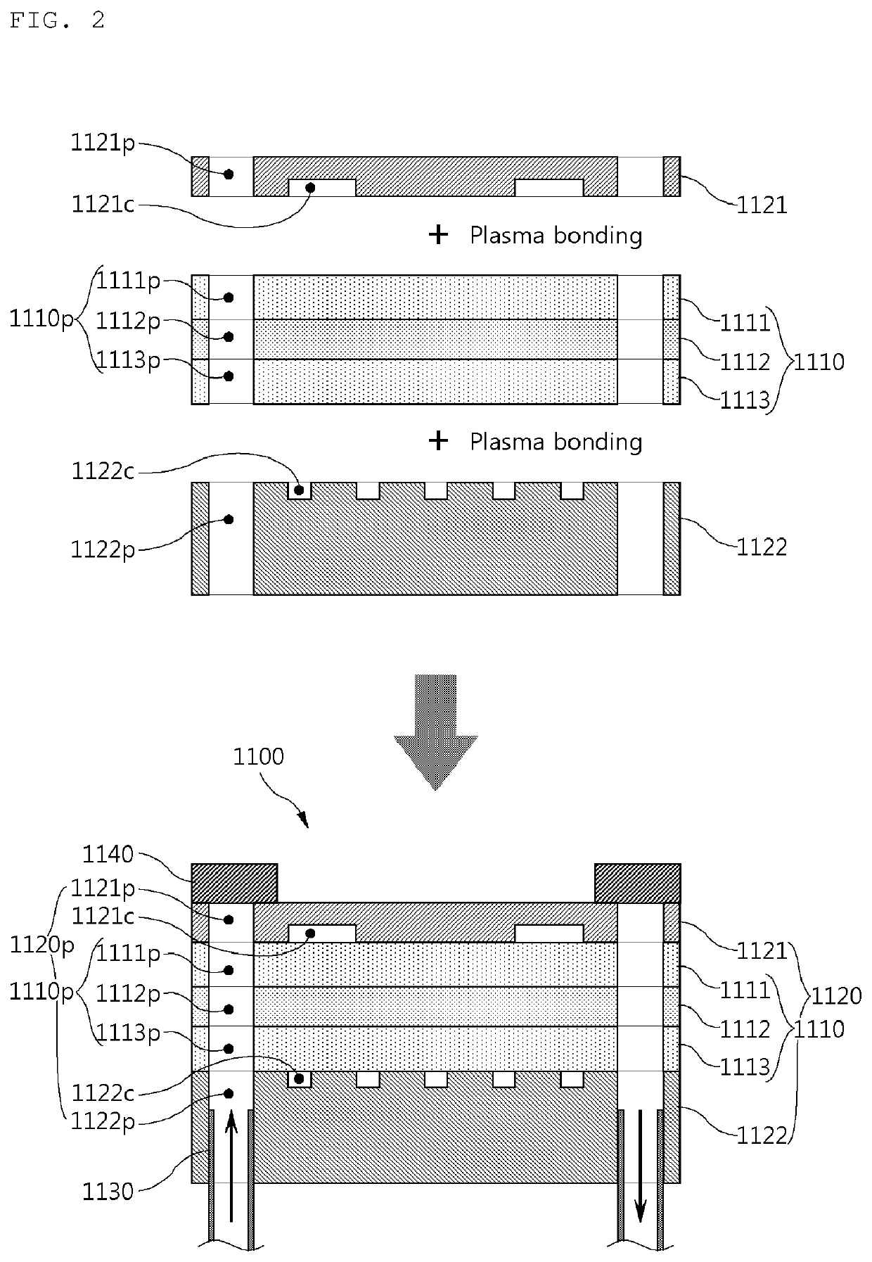
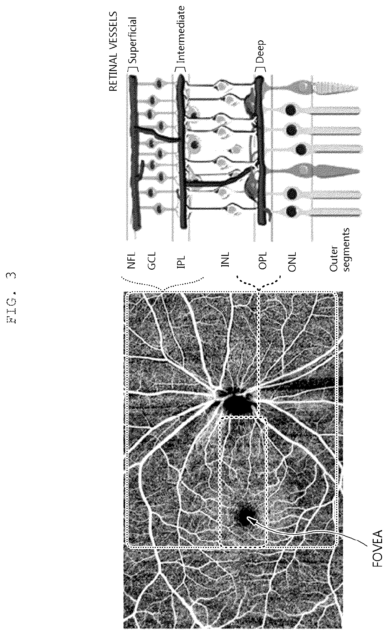
]1000: Eye phantom1100: Retina simulating part1110: Multilayer film structure part1110p: Multilayer filmflow passage part1111: GCL simulating part1111p: GCL flow passage part1112: IPL simulating part1112p: IPL flow passage part1113: INL simulating part1113p: INL flow passage part1120: Vascular layer structure part1120p: Blood vessel layer1121: NFL simulating partflow passage part1121c: NFL channel part1121p: NFL flow passage part1122: OPL simulating part1122c: OPL channel part1122p: OPL flow passage part1130: Flow passage part1140: Sealing part1150: Outer film structure part1151: ONL simulating part1152: Outer segmentsimulating part1200: Lens part1300: Housing part

BEST MODE

[0043]Hereinafter, an eye phantom for evaluating a retinal angiography image and a manufacturing method therefor according to the present invention having the above-described configuration will be described in detail with reference to the accompanying drawings.

[0044]Retina Simulating Part Structure and Manufacturi...


PUM


Abstract

The present invention relates to an eye phantom for evaluating a retinal angiography image, and a manufacturing method therefor. A purpose of the present invention is to provide an eye phantom for evaluating a retinal angiography image, capable of simulating even the vascular structure and blood flow of a retina so as to be more similar to an actual retinal structure than a conventional eye phantom; and a manufacturing method therefor. More specifically, a purpose of the present invention is to provide an eye phantom for evaluating a retinal angiography image, including various shapes of fine fluid channel structures corresponding to the vascular structure formed in an actual retina; and a manufacturing method therefor.

Description

CROSS-REFERENCE TO RELATED APPLICATIONS[0001]This application is the United States national phase of International Application No. PCT / KR2019 / 013198 filed Oct. 8, 2019, and claims priority to Korean Patent Application No. 10-2019-0008743 filed Jan. 23, 2019, the disclosures of which are hereby incorporated by reference in their entirety.BACKGROUND OF THE INVENTIONField of the Invention[0002]The present invention relates to an eye phantom for evaluating a retinal angiography image, and more particularly, to an eye phantom for evaluating a retinal angiography image which is used to evaluate an optical coherence tomography (OCT) for ophthalmology that photographs an image to observe a retina of an eyeball or the like, and can more easily and smoothly perform a performance evaluation of the OCT by simulating an actual structure of the eyeball more similarly.Description of Related Art[0003]A retina of a human eye is a core tissue that detects light and determines vision, and is a very th...

Claims


Application Information

Patent Timeline
IPC IPC(8): A61B3/00A61B3/12A61B3/10
CPCA61B3/0025A61B3/102A61B3/1241G09B23/286G09B23/303A61B3/00
Inventor LEE, SANG WONDOH, ILLEE, HYUNJI
Owner KOREA RES INST OF STANDARDS & SCI
Learn More