
Automatic pleural-peritonal pump

a pleural cavity and peritonal pump technology, applied in the direction of wound drains, process and machine control, instruments, etc., can solve the problems of pathological compression of lung tissue, unbalanced net flow of pleural fluid within the pleural cavity, and considerable difficulty in or prevention of breathing process, so as to avoid infection and other complications, relieve symptoms, and high success rate of treatmen

Active Publication Date: 2021-09-16
PLEURAL DYNAMICS INC
View PDF7 Cites 2 Cited by
  • Summary
  • Abstract
  • Description
  • Claims
  • Application Information

AI Technical Summary

Benefits of technology

This patent text describes a new way to drain excess fluid from the chest (pleural effusions) that reduces the risk of infection and other complications. The technique involves using an automatic pump that can be implanted in the patient and managed by a caregiver. This pump uses a special catheter that is designed to drain fluid regularly and without the need for manual compression or an external reservoir. The use of the automatic pump also reduces the risk of clotting and allows for improved performance. Overall, this new technique provides a more efficient and effective way to manage fluid buildup in the chest.

Problems solved by technology

However, under certain abnormal conditions, such as infection, inflammation, malignancy, heart failure, liver failure, or kidney failure, among other conditions, the net flow of pleural fluid within the pleural cavity becomes unbalanced, with increased fluid production, decreased reabsorption, or both, resulting in the excess accumulation (e.g., on the order several hundred milliliters to several liters) of fluid in the pleural space.
Excess accumulation of pleural fluid is known as pleural effusion and adds additional mass that must be moved with each breath and may cause the pathological compression of lung tissue.
This results in considerable difficulty in or prevention of the breathing process.
Pleural effusion may lead to, for example, dyspnea, shortness of breath, chest pain, and / or chronic cough, and may greatly compromise a patient's quality of life.
Many of these effusions are chronic, recur if drained, and can be quite symptomatic and debilitating for the patient.
Unfortunately, malignant effusions are likely to recur following drainage and in order to control symptoms therapeutic thoracentesis must be repeated frequently.
However, due to patients' delays in notifying their physician that their symptoms have returned and delays in organizing and providing repeat thoracentesis, patients often spend a significant portion of their life with effusion-related symptoms.
Furthermore, thoracentesis is painful and uncomfortable, and is frequently accompanied by complications such as pneumothorax (i.e., the collapse of the lung due to accumulation of air in the pleural cavity) in up to eleven percent of patients and severe bleeding or infection in many others.
Unfortunately, chest-tube pleurodesis typically requires hospitalization for at least two days and as many as seven days, can be quite painful, can lead to breathing difficulties related to the pleurodesis, and, in up to one third of patients, fails to provide relief of symptoms for more than a few weeks.
Unfortunately, thoracoscopic pleurodesis typically still requires prolonged hospitalization for four to seven days, can also be quite painful, can lead to breathing difficulties related to the pleurodesis, and, in many patients, fails to provide relief of symptoms for more than a few weeks.
However, because the catheter passes through the patient's skin with a portion inside the body and a portion outside the body, a considerable fraction of patients, approximately eight percent, fall victim to infection.
Further, the patient is subject to the discomfort, irritation, and annoyance of an exposed indwelling catheter.
However, also like chronic indwelling catheters, a considerable percentage of patients, approximately four percent, fall victim to infection.
Further drawbacks of conventional pleuroperitoneal shunts include a relatively high rate of shunt-specific complications, such as clotting of fluid within the shunt.
Finally, in order to gain relief from effusion related symptoms, the patient or their caregiver must actively compress the pumping chamber multiple repeatedly to transfer effusion fluid from the pleural space to the peritoneal space causing significant discomfort and inconvenience.

Method used


Image

  • Automatic pleural-peritonal pump
  • Automatic pleural-peritonal pump
  • Automatic pleural-peritonal pump

Examples


Embodiment Construction

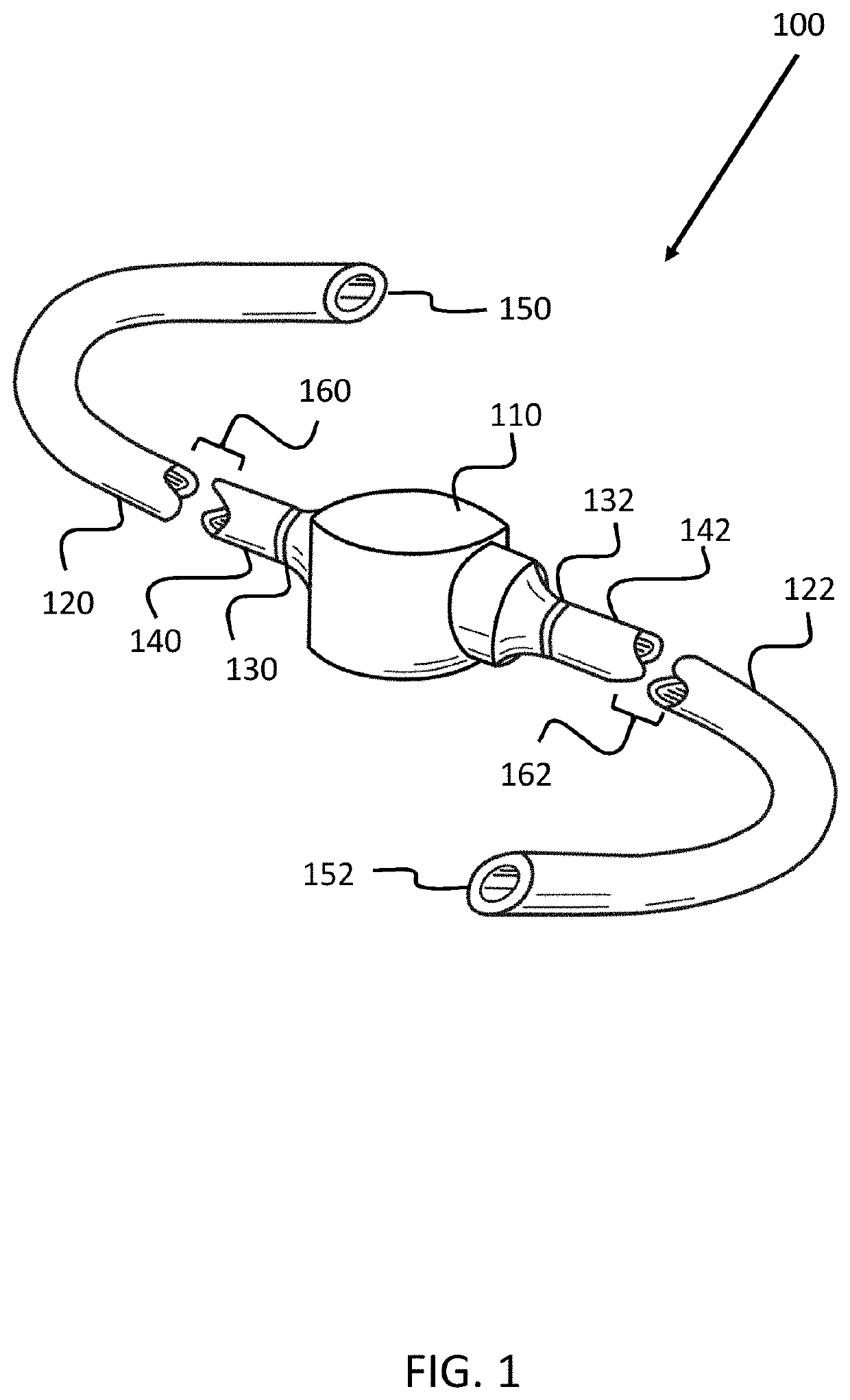
[0056]The apparatuses, systems, and methods described herein may be used for the purposes of draining and / or moving fluid from one cavity within the human body to another cavity. Particularly, the apparatuses, systems, and methods described herein comprise an automatic pump which provides a general pumping function in an automatic pump-based fluid management system.

[0057]For purposes of explanation, the disclosure herein includes a discussion of the use of an automatic pump-based fluid management system for the purposes of drainage of pleural fluid for the treatment of pleural effusions. However, it should be understood that such an application is but one particular application of one particular embodiment of an automatic pump-based fluid management system, and that other embodiments and applications are possible.

[0058]Also, for purposes of explanation, the disclosure herein describes an automatic pump as part of a particular automatic pump-based fluid management systems. However, i...


PUM


Abstract

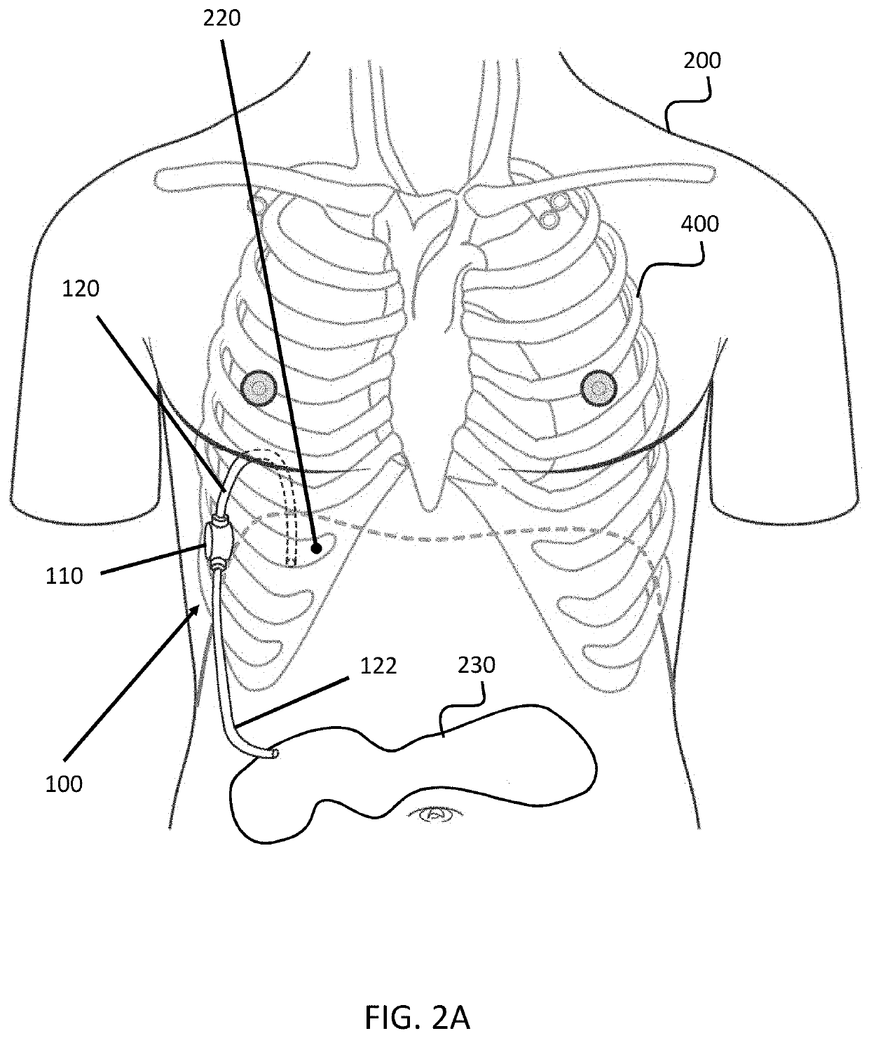
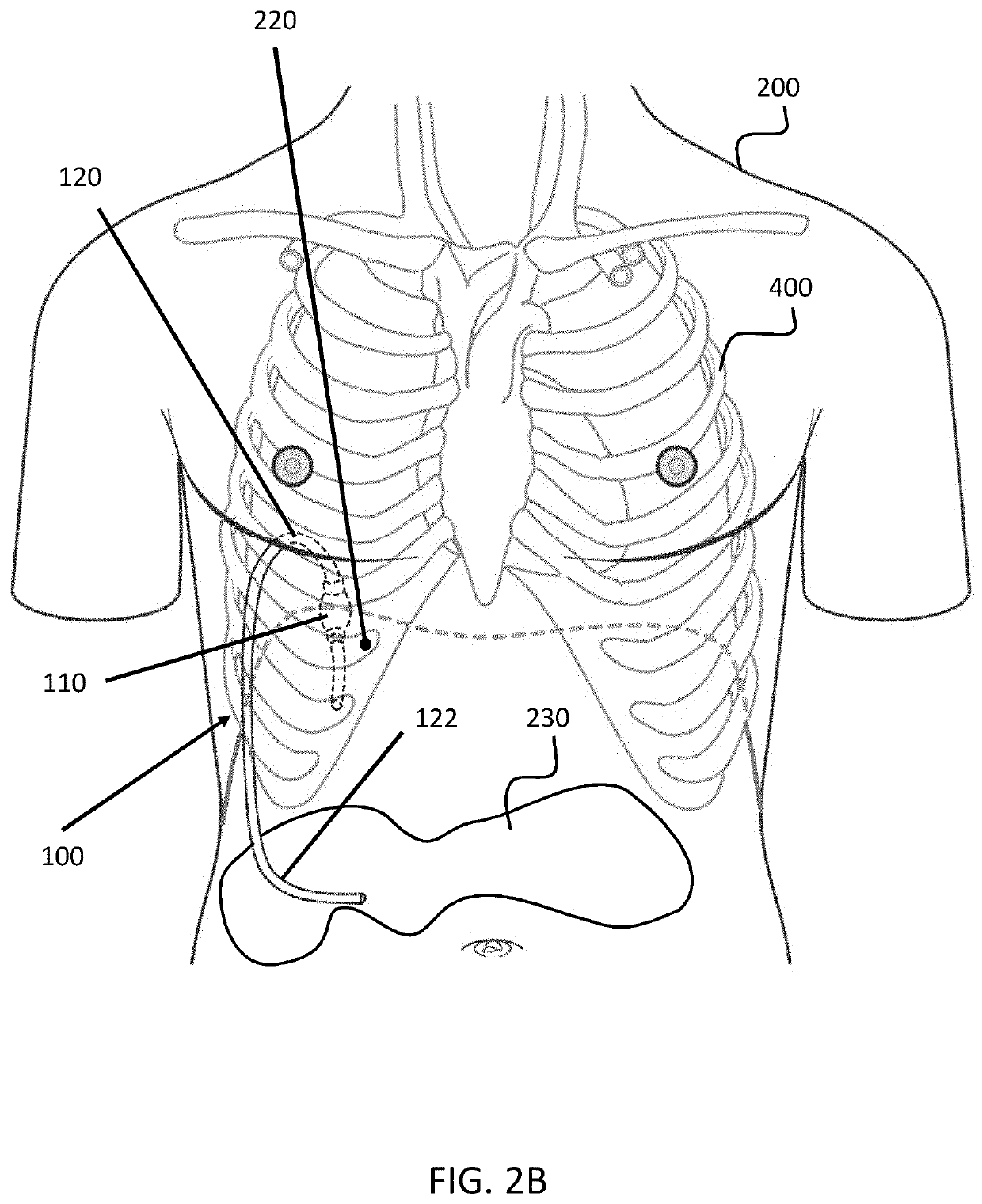
An automatic pump-based fluid management system, as described herein, comprises an intercostal pump that is, generally, a resiliently flexible bulb having an inlet and an outlet. The inlet is attached to a first tube that extends from the intercostal pump to a first area of a patient's body, for example, the patient's pleural cavity. The outlet is connected to a second tube that extends from the intercostal pump to a second area of a patient's body, for example, the patient's peritoneal cavity. In use, the intercostal pump is placed between a first rib and a second rib in a patient. The intercostal pump operates by being successively compressed and decompressed between the first and second ribs as the patient breaths.

Description

RELATED APPLICATIONS[0001]This application relates to U.S. patent application. Ser. No. 13 / 968,479, filed on Aug. 16, 2013, entitled “Systems and Methods for Draining Bodily Fluid via an Intercostal Pump,” which is hereby incorporated by reference herein in its entirety.BACKGROUND[0002]A number of techniques for draining bodily fluid involve utilizing a pump, in combination with a shunt or catheter, to drain fluid from one cavity within the human body to either another cavity or to a reservoir outside of the body. Such techniques may be utilized for purposes including, for example, draining a patient's blood, urine, saliva, cerebrospinal fluid, peritoneal fluid, and / or pleural fluid, among other possibilities.[0003]One drainage-technique application is the drainage of pleural fluid for the treatment of pleural effusions. Pleural fluid is normally a low-protein liquid that can be found in relatively small amounts (normally 10-20 milliliters) in each of a patient's pleural cavities. T...

Claims


Application Information

Patent Timeline
Patent Type & Authority Applications(United States)
IPC IPC(8): A61M5/142A61M39/24A61M5/168
CPCA61M5/14276A61M39/24A61M5/16804A61M2205/3334A61M2205/8206A61M2205/3379A61M2039/2406A61M1/82A61M1/84A61M1/71A61M27/002A61M2210/101A61M2210/1017
Inventor MAYSE, MARTIN L.
Owner PLEURAL DYNAMICS INC
Learn More