
Deep learning based medical image detection method and related device

a deep learning and medical image technology, applied in the field of artificial intelligence (ai), can solve the problems of low reliability of medical image detection models, loss of information of 3d volume data of ct images, and general only suitable, so as to improve the prediction reliability of a region of interes

Pending Publication Date: 2021-08-26
TENCENT TECH (SHENZHEN) CO LTD
View PDF0 Cites 6 Cited by
  • Summary
  • Abstract
  • Description
  • Claims
  • Application Information

AI Technical Summary

Benefits of technology

The patent text describes a deep learning-based method and apparatus for detecting regions of interest in medical images. The method uses a deep neural network to process each slice of the image and predict the position and confidence score of the region of interest. The method also merges feature maps from different layers of the image to detect objects of different scales. The method can be trained using a relatively small amount of data and is efficient in detecting the position and confidence score of the region of interest in the image. The technical effects of the patent text include improving the accuracy and reliability of predicting the position and confidence score of regions of interest in medical images and assisting doctors in detecting suspected lesion regions in multi-scale CT images.

Problems solved by technology

This method is generally only suitable for image data such as mammary gland.
In a case that a Computed Tomography (CT) image is processed using the method, information of 3D volume data of the CT image would be lost, and the reliability of the medical image detection model is relatively low.
Hence, in the field of medical image detection, how to comprehensively use the 3D volume data of the medical image to improve the reliability of region-of-interest prediction is a technical problem that needs to be resolved urgently.

Method used


Image

  • Deep learning based medical image detection method and related device
  • Deep learning based medical image detection method and related device
  • Deep learning based medical image detection method and related device

Examples


Embodiment Construction

[0039]At present, the exemplary implementations are described comprehensively with reference to the accompanying drawings. However, the examples of implementations may be implemented in a plurality of forms, and it is not to be understood as being limited to the examples described herein. Conversely, the implementations are provided to make this application more comprehensive and complete, and comprehensively convey the idea of the examples of the implementations to a person skilled in the art.

[0040]In addition, the described features, structures or characteristics may be combined in one or more embodiments in any appropriate manner. In the following descriptions, a lot of specific details are provided to give a comprehensive understanding of the embodiments of this application. However, a person of ordinary skill in the art is to be aware that, the technical solutions in this application may be implemented without one or more of the particular details, or another method, unit, appa...


PUM


Abstract

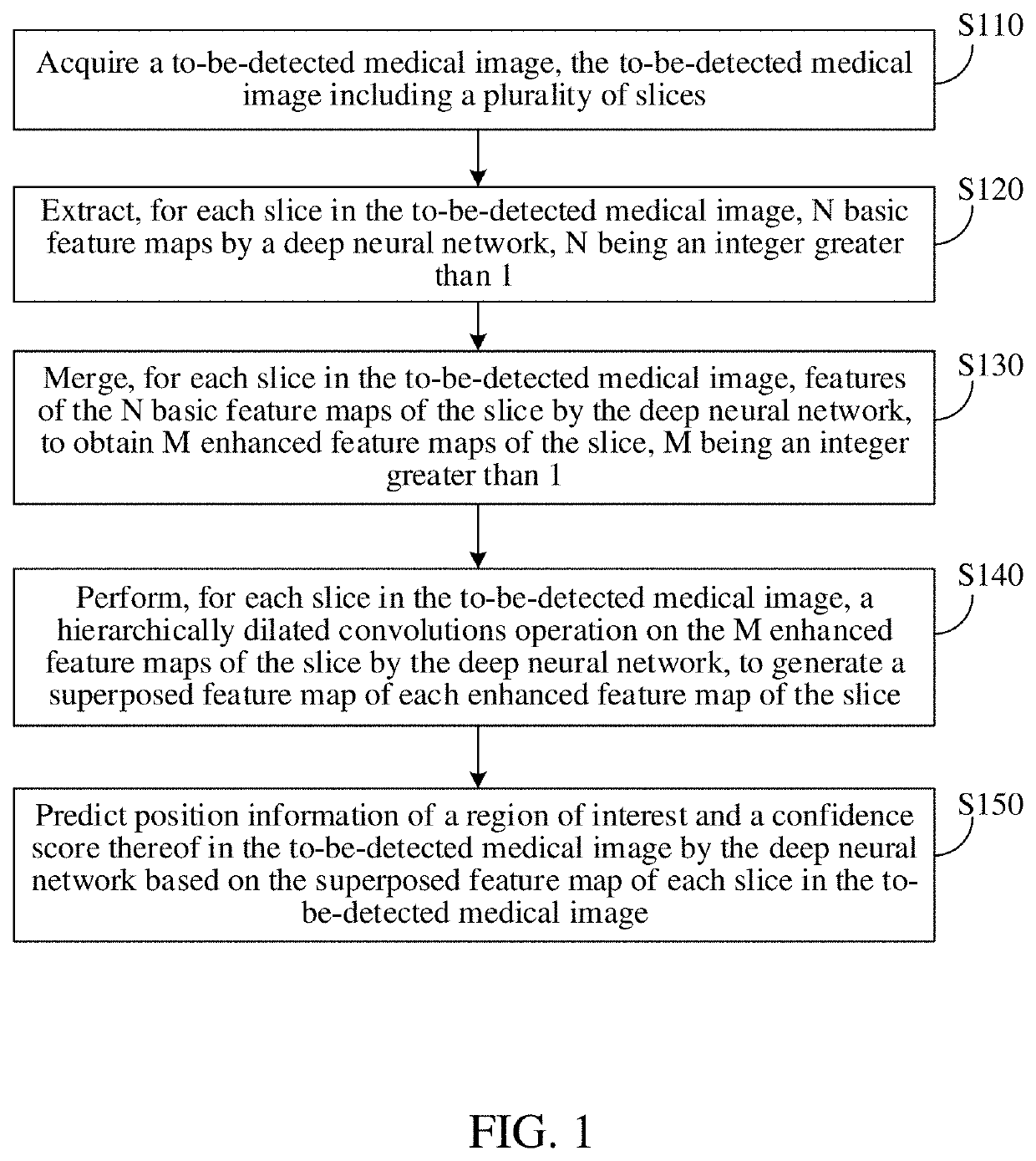
The present disclosure provides a deep learning based medical image detection method and apparatus, a computer-readable medium, and an electronic device. The method includes: acquiring a to-be-detected medical image comprising a plurality of slices; for each slice in the to-be-detected medical image: extracting N basic feature maps of the slice by a deep neural network, N being an integer greater than 1, merging features of the N basic feature maps by the deep neural network, to obtain M enhanced feature maps, M being an integer greater than 1, and respectively performing a hierarchically dilated convolutions operation on the M enhanced feature maps by the deep neural network, to generate a superposed feature map of each enhanced feature map; and predicting position information of a region of interest and a confidence score thereof in the to-be-detected medical image by the deep neural network based on the superposed feature map.

Description

RELATED APPLICATION[0001]This application is a continuation application of PCT Patent Application No. PCT / CN2020 / 081655, filed on Mar. 27, 2020, which claims priority to Chinese Patent Application No. 201910324565.8 filed with the China National Intellectual Property Administration on Apr. 22, 2019, both of which are incorporated herein by reference in their entireties.FIELD OF THE TECHNOLOGY[0002]This application relates to the technical field of Artificial Intelligence (AI), and in particular, to a medical image detection technique.BACKGROUND OF THE DISCLOSURE[0003]At present, a medical image detection model is mainly adopted to perform image detection on data of a two-dimension (2D) image, to recognize a region of interest in the 2D image. This method is generally only suitable for image data such as mammary gland. In a case that a Computed Tomography (CT) image is processed using the method, information of 3D volume data of the CT image would be lost, and the reliability of the ...

Claims


Application Information

Patent Timeline
Patent Type & Authority Applications(United States)
IPC IPC(8): G06T7/00G06K9/32G06T3/40G06V10/25
CPCG06T7/0012G06K9/3241G06T2207/20084G06T2207/20081G06T3/4053G06T2207/10081G06T2207/20016G06T2207/30096G06T2207/20221G06F18/253G06V10/25G06V2201/03G06N3/084G06V10/82G06V10/806G06N3/045
Inventor GONG, LIJUN
Owner TENCENT TECH (SHENZHEN) CO LTD
Learn More