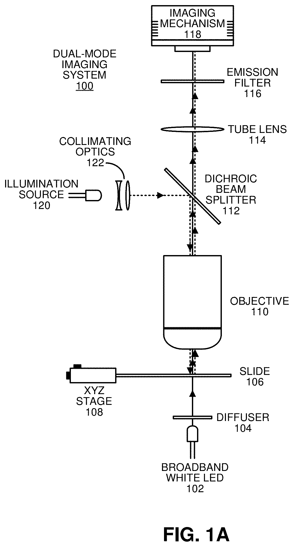
Producing a composite image of a stained tissue sample by combining image data obtained through brightfield and fluorescence imaging modes

a tissue sample and composite image technology, applied in the field of tissue sample composite image production, can solve the problems of increasing cost, creating workflow complications, and underestimating collagen content of trichrome stains, and achieve the effect of facilitating toggling

Pending Publication Date: 2021-07-01
RGT UNIV OF CALIFORNIA
View PDF7 Cites 2 Cited by
  • Summary
  • Abstract
  • Description
  • Claims
  • Application Information

AI Technical Summary

Benefits of technology

[0010]In some embodiments, while processing the image data, the system performs non-component-specific image-processing operations on the image data to improve image quality.
[0013]In some embodiments, while processing the image data, the system performs image-processing operations on the fluorescence image data to improve image quality. Next, while combining the image data, the system combines the processed fluorescence image data with the brightfield histology image to generate the composite image, which provides more information than the brightfield histology image alone.
[0030]In some embodiments, the system displays the composite image through a display system that facilitates toggling among two or more of the composite image, the brightfield histology image, the fluorescence image, and an extracted targeted component image.

Problems solved by technology

However, widely used traditional trichrome stains have been found to underestimate collagen content.
However, it is not routinely used in clinical histology, and also involves an extra slide and staining step, which increases cost and creates workflow complications and can be problematic for very small specimens.
However, SHG is an expensive approach, which requires multi-photon lasers and confocal scanning optics, and is specific to non-centrosymmetric molecules such as collagens I, II, and III and is also highly orientation-dependent.
Moreover, generating strong, detectable SHG signals requires some degree of alignment between light polarization and collagen fiber direction and this technique is unable to highlight collagen type IV.

Method used


Image

  • Producing a composite image of a stained tissue sample by combining image data obtained through brightfield and fluorescence imaging modes
  • Producing a composite image of a stained tissue sample by combining image data obtained through brightfield and fluorescence imaging modes
  • Producing a composite image of a stained tissue sample by combining image data obtained through brightfield and fluorescence imaging modes

Examples


Embodiment Construction

[0040]The following description is presented to enable any person skilled in the art to make and use the present embodiments, and is provided in the context of a particular application and its requirements. Various modifications to the disclosed embodiments will be readily apparent to those skilled in the art, and the general principles defined herein may be applied to other embodiments and applications without departing from the spirit and scope of the present embodiments. Thus, the present embodiments are not limited to the embodiments shown, but are to be accorded the widest scope consistent with the principles and features disclosed herein.

[0041]The data structures and code described in this detailed description are typically stored on a computer-readable storage medium, which may be any device or medium that can store code and / or data for use by a computer system. The computer-readable storage medium includes, but is not limited to, volatile memory, non-volatile memory, magneti...


PUM

PropertyMeasurementUnit
fluorescence imagingaaaaaaaaaa
brightfield imagingaaaaaaaaaa
multispectral imaging systemaaaaaaaaaa

Abstract

The disclosed embodiments relate to a system that produces a composite image of a stained tissue sample by combining image data obtained through brightfield and fluorescence imaging modes. While operating in a brightfield imaging mode, the system illuminates the stained tissue sample with broadband light, and collects image data comprising a brightfield histology image using a multispectral imaging system. While operating in a fluorescence imaging mode, the system illuminates the stained tissue sample with one or more bands of excitation light, and collects image data associated with resulting fluorescence emissions using the multispectral imaging system. Next, the system processes the image data collected during the brightfield and / or fluorescence imaging modes. Finally, the system combines the image data collected during the brightfield and fluorescence imaging modes to produce the composite image.

Description

RELATED APPLICATION[0001]This application claims priority under 35 U.S.C. § 119 to U.S. Provisional Application No. 62 / 691,095, entitled “Detecting the Spatial Distribution of Collagen and other Tissue Structural Components on H & E Slides without Additional Stains or Complicated Optics” by the same inventors as the instant application, filed on 28 Jun. 2018, the contents of which are incorporated by reference herein.BACKGROUNDField[0002]The disclosed embodiments generally relate to techniques for producing images of tissue samples. More specifically, the disclosed embodiments relate to a technique for producing a composite image of a stained tissue sample by combining image data obtained through brightfield and fluorescence imaging modes, wherein the composite image can be used to better visualize structural macromolecule-related tissue components, such as components comprised of collagen.Related Art[0003]Collagen is a major component of the extracellular matrix, which in the tumor...

Claims


Application Information

Patent Timeline
Patent Type & Authority Applications(United States)
IPC IPC(8): G01N21/64G01N1/30G02B21/00G06V10/143
CPCG01N21/6428G01N21/6458G01N2021/6441G02B21/0076G01N1/30G02B21/06G02B21/36G02B21/367G02B21/0032G01J3/4406G01J3/51G01J2003/2826G01J3/2823G06V20/69G06V10/143G06V2201/03G01J3/10
Inventor FEREIDOUNI, FARZADLEVENSON, RICHARD M.
Owner RGT UNIV OF CALIFORNIA
Why Eureka
Try Eureka