
Method of Operation of An Artificial Intelligence-Equipped Specimen Scanning and Analysis Unit to Digitally Scan and Analyze Pathological Specimen Slides

a technology of artificial intelligence and specimen scanning, applied in the field of digital pathology, can solve the problems of restricted market market of digital pathology slide scanners today

Active Publication Date: 2020-10-22
OPTRASCAN INC
View PDF0 Cites 0 Cited by
  • Summary
  • Abstract
  • Description
  • Claims
  • Application Information

AI Technical Summary

Benefits of technology

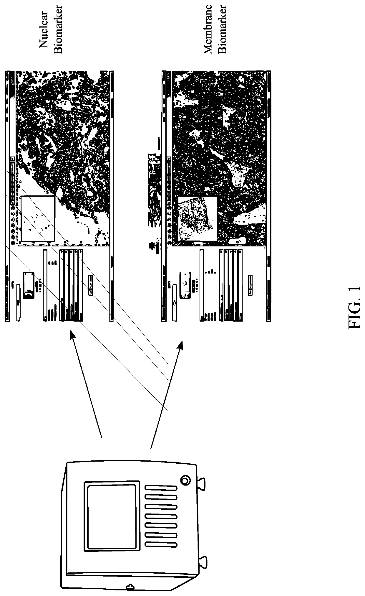
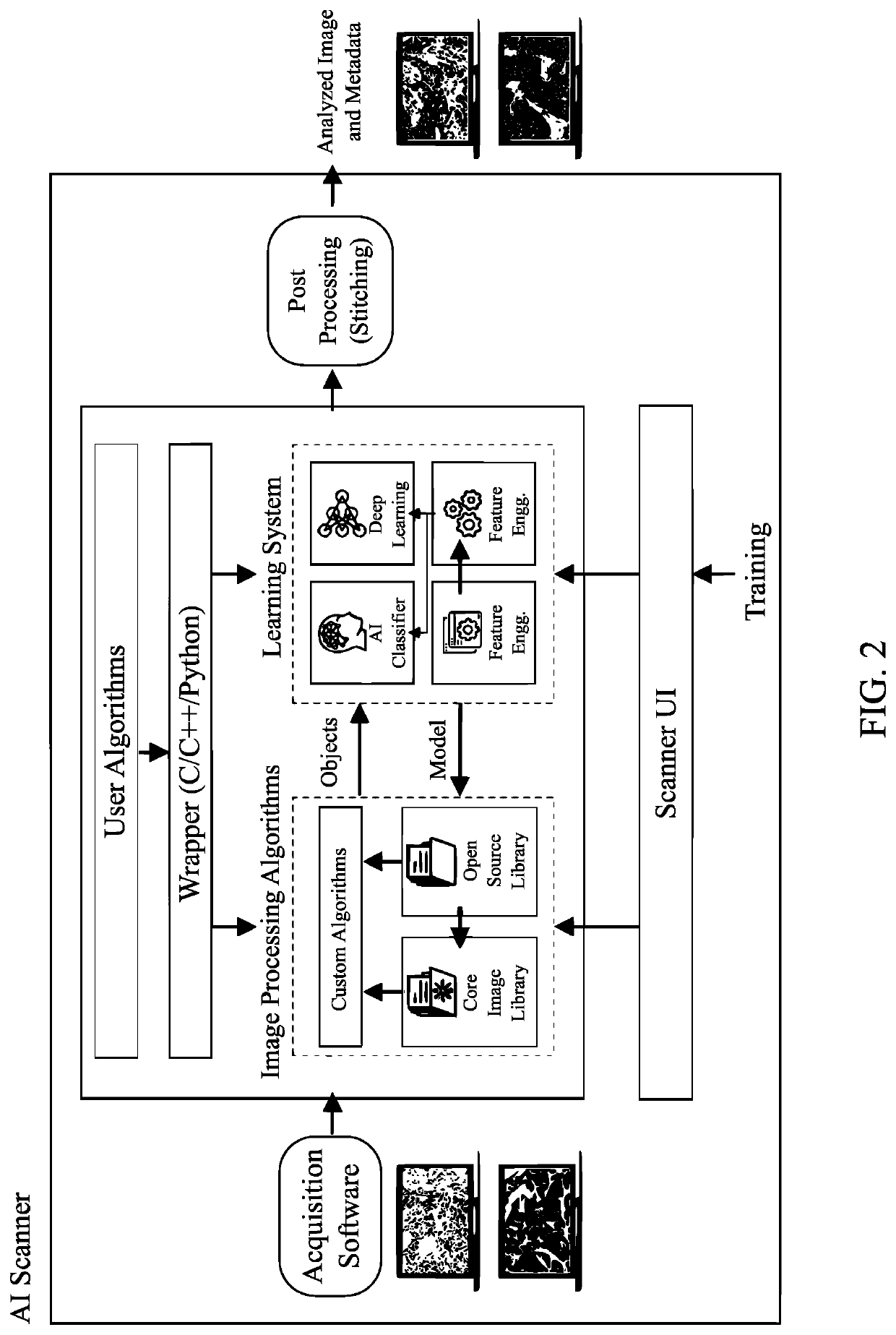
The present invention is a new generation of artificial intelligence (AIvention) that is enabled by a digital pathology slide scanning unit. This scanning unit is capable of not only scanning slides but also performing digital pathological analysis on the scanned images for the purposes of immunopathology. The scanning unit provides a benefit to histopathologists and cytopathologists as they can utilize a single unit to not only scan a slide but also view the results of a pathological analysis of the tissue sample mounted to the slide. The invention utilizes a custom algorithm that has one or more embedded AI frameworks and plug-in architecture to process captured images. The custom algorithms provide features such as real-time region of interest detection, cell quantification, rare event detection, and morphological and image data structure manipulation algorithms. The invention aims to improve the efficiency and accuracy of digital pathology analysis and diagnosis.

Problems solved by technology

Digital Pathology slide scanners today in the market are restricted to partial or whole slide image acquisition and digitization of slide into an image.

Method used


Image

  • Method of Operation of An Artificial Intelligence-Equipped Specimen Scanning and Analysis Unit to Digitally Scan and Analyze Pathological Specimen Slides
  • Method of Operation of An Artificial Intelligence-Equipped Specimen Scanning and Analysis Unit to Digitally Scan and Analyze Pathological Specimen Slides
  • Method of Operation of An Artificial Intelligence-Equipped Specimen Scanning and Analysis Unit to Digitally Scan and Analyze Pathological Specimen Slides

Examples


Embodiment Construction

[0015]All illustrations of the drawings are for the purpose of describing selected versions of the present invention and are not intended to limit the scope of the present invention. The present invention is to be described in detail and is provided in a manner that establishes a thorough understanding of the present invention. There may be aspects of the present invention that may be practiced or utilized without the implementation of some features as they are described. It should be understood that some details have not been described in detail in order to not unnecessarily obscure focus of the invention. References herein to “the preferred embodiment”, “one embodiment”, “some embodiments”, or “alternative embodiments” should be considered to be illustrating aspects of the present invention that may potentially vary in some instances, and should not be considered to be limiting to the scope of the present invention as a whole.

[0016]The present invention relates to a new generation...


PUM


Abstract

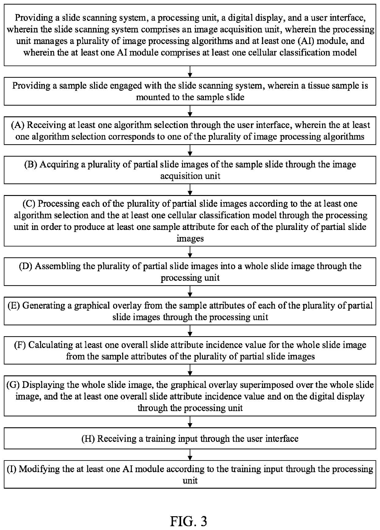
In a method of operation of an artificial intelligence-equipped specimen scanning and analysis unit to digitally scan and analyze pathological specimen slides, a sample slide with a mounted tissue sample is scanned and analyzed according to one or more user-selected algorithms in order to generate a heatmap visually depicting the presence of one or more user-selected sample attributes of the tissue sample. One or more artificial intelligence modules, including a deep learning computation module, is provided and can be trained by the user for future analysis of new samples. One or more regions of interest may be selected from the heatmap to include in the results of the analysis. A focus window may be used to closely inspect any given region of the whole slide image, and a trail map is generated from the movement of the focus window.

Description

[0001]The current application claims a priority to the U.S. Provisional Patent application Ser. No. 62 / 836,271 filed on Apr. 19, 2019. The current application is filed on Apr. 20, 2019, whereas Apr. 19, 2019 was on a weekend.FIELD OF THE INVENTION[0002]The present invention relates generally to digital pathology. More particularly, the present invention relates to image acquisition and analysis of biological specimens in a singular, self-contained unit.BACKGROUND OF THE INVENTION[0003]Digital Pathology slide scanners today in the market are restricted to partial or whole slide image acquisition and digitization of slide into an image. There exists a demand for a singular unit capable of scanning and analyzing a tissue sample on a sample slide.[0004]Additional advantages of the invention will be set forth in part in the description which follows, and in part will be obvious from the description, or may be learned by practice of the invention. Additional advantages of the invention ma...

Claims


Application Information

Patent Timeline
IPC IPC(8): G06T7/00G06K9/62G06N20/00
CPCG06T2207/20212G06T2207/20081G06T7/0014G06N20/00G06T2207/30096G06K9/6268G06T2207/30024G06K9/6228G06T2207/10056G06N3/08G06V20/698G06V10/235G06F18/211G06F18/241
Inventor GHOLAP, ABHIJEET SHARADCHANDRADOSHI, ISHAKAMBLE, GURUNATH
Owner OPTRASCAN INC
Learn More