Eureka AIR delivers breakthrough ideas for toughest innovation challenges, trusted by R&D personnel around the world.

Computer-implemented machine learning for detection and statistical analysis of errors by healthcare providers

a technology of machine learning and error detection, applied in the field of computer-implemented machine learning for diagnostic error detection, can solve problems such as differences in communication, complex interpretation of digital images, and preparation for or performing surgical procedures that are unnecessary

Active Publication Date: 2020-10-22
COVERA HEALTH
View PDF0 Cites 28 Cited by
  • Summary
  • Abstract
  • Description
  • Claims
  • Application Information

AI Technical Summary

Benefits of technology

The present invention provides systems and methods for training a machine learning network for diagnostic quality assessment. The method involves training a network using two encoder networks, one for text and one for images, and a concept generator. The network is trained using a combination of binary cross entropy loss and categorical cross entropy loss between the training data and labeled data. The resulting network can accurately assess the quality of diagnostic images and text. The technical effect of this invention is to improve the accuracy and efficiency of diagnostic quality assessment.

Problems solved by technology

However, the interpretation of digital images is often complex, requiring significant medical and anatomical knowledge as well as an ability to detect subtle or complicated patterns of information in the correct context, and therefore the radiology field has a non-zero error rate, in which patients have had their diagnostic image data interpreted incorrectly, leading to the wrong diagnosis.
For example, an erroneous diagnosis could lead to preparation for or performance of a surgical procedure that is unnecessary.
Some diagnostic errors result from deficiencies in a radiologist's skill in interpreting image data, other diagnostic errors result from differences in the communication of diagnostic information in written or dictated diagnostic reports.
A wide variety of diagnostic errors and quality issues occur with varying prevalence rates in patient exams.
Other quality issues, related to communication issues in the report, can include the following categories: (1) findings that are reported in an overly equivocal manner, (2) findings that are reported in an overly vague manner, (3) findings that are reported with inappropriate emphasis, (4) inappropriate or lack of comparisons with prior diagnostic studies, (5) inappropriate or lack of inclusion of relevant standard measures (e.g. not using the Breast Imaging Reporting and Data System or BI-RADS scoring system for mammogram reports), or (6) inappropriate or lack of follow-up recommendations.
Finally, diagnostic radiology exams can also suffer from technical errors and quality issues that can include: (1) poor image quality (e.g. low signal-to-noise ratio), (2) images degraded or obscured by patient motion or other artifacts, (3) poorly configured exam protocols (e.g. an MRI exam conducted without collecting images that have a necessary image contrast setting or images collected with resolution that is too low), or (4) poor anatomical coverage of the images.
Assessing the accuracy of diagnoses and presence of specific types of errors is difficult for patients and other stakeholders, including other physicians involved in a patient's care and healthcare payers.
While a diagnostic accuracy assessment could be based upon favoring the second opinion of an authoritative expert, the healthcare system might not be well-served if correct diagnoses only can be achieved by a subset of experts.
Furthermore, authoritative experts are themselves fallible and pathological assessment always involves a measure of subjectivity, so it may be difficult to determine if variation across the two diagnoses represent evidence of diagnostic errors present in at least one diagnosis or if the variation represents multiple ways of stating the same diagnosis.
Seeking a third or multiple additional opinions on a given patient's diagnosis does not alleviate this issue and is likely prohibitive due to logistics or cost for most patients.
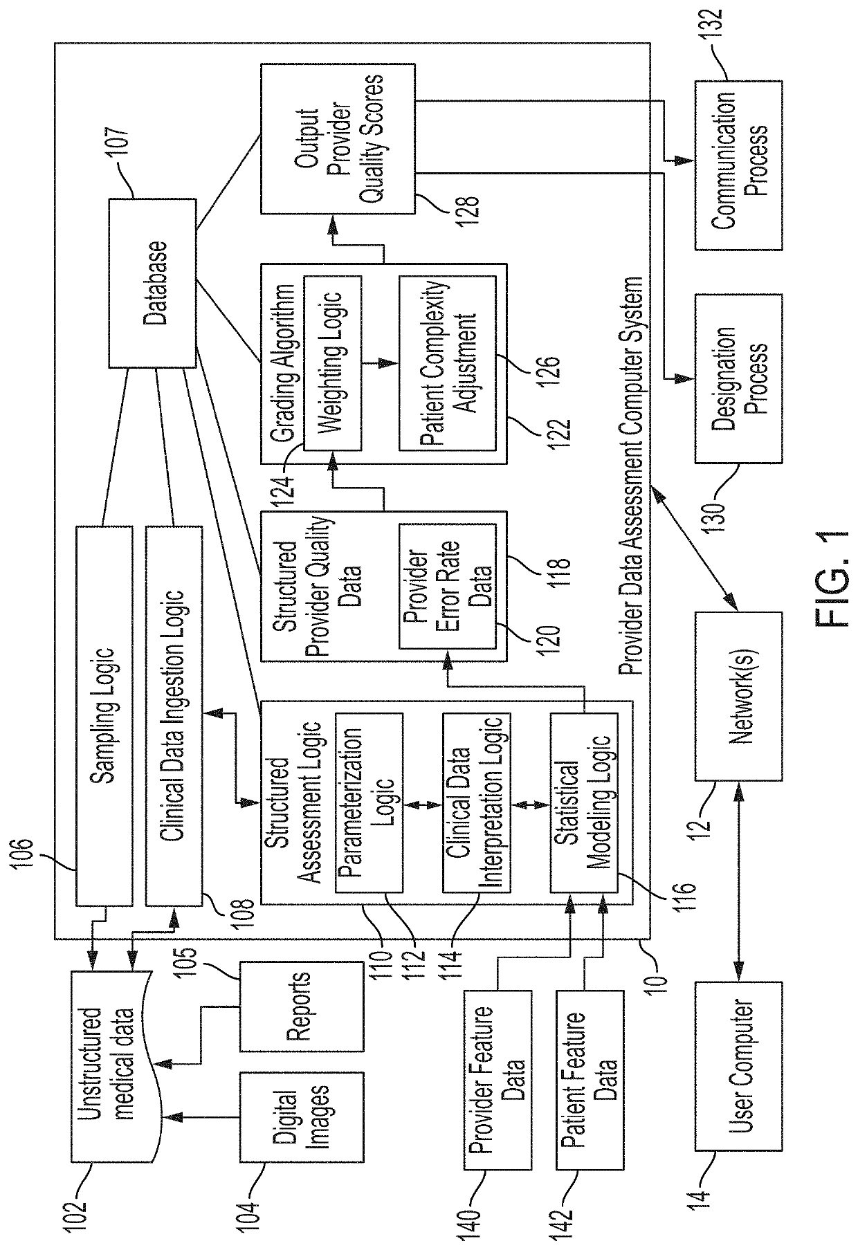
While extensive medical records are usually developed for each patient in digital electronic form, typically much of the data is unstructured; examples are the digital medical images and dictated diagnostic reports, both of which are non-standardized across patient exams and not readily interpretable by machines or computers.
While more structured dictation could be provided, it is an imperfect approach that is unlikely to be adopted on a widespread basis.
However, if estimates of accuracy rates and error rates are directly based on data generated by independent radiologists who use a standardized process for identifying and characterizing selected diagnostic findings and diagnostic errors, the estimates will themselves not be accurate or reliable due to inter-radiologist variability.
However, since not all diagnostic errors and quality issues have the same impact on downstream patient care patterns or patient outcomes, straightforward estimates of diagnostic accuracy rates or error rates may not represent a valuable quality metric.
It may be inappropriate to use unadjusted estimates of diagnostic accuracy rates or error rates as standardized and generalizable measures of radiology care quality.
However, known systems typically identify too many false positives, or work only with abnormalities that are straightforward to find in an image, and therefore they do not add significant value to the ecosystem in this capacity.
Computer-implemented image interpretation and medical report interpretation technologies have not been developed, expanded, or adapted for use as part of a diagnostic accuracy and quality assessment system.

Method used


Image

  • Computer-implemented machine learning for detection and statistical analysis of errors by healthcare providers
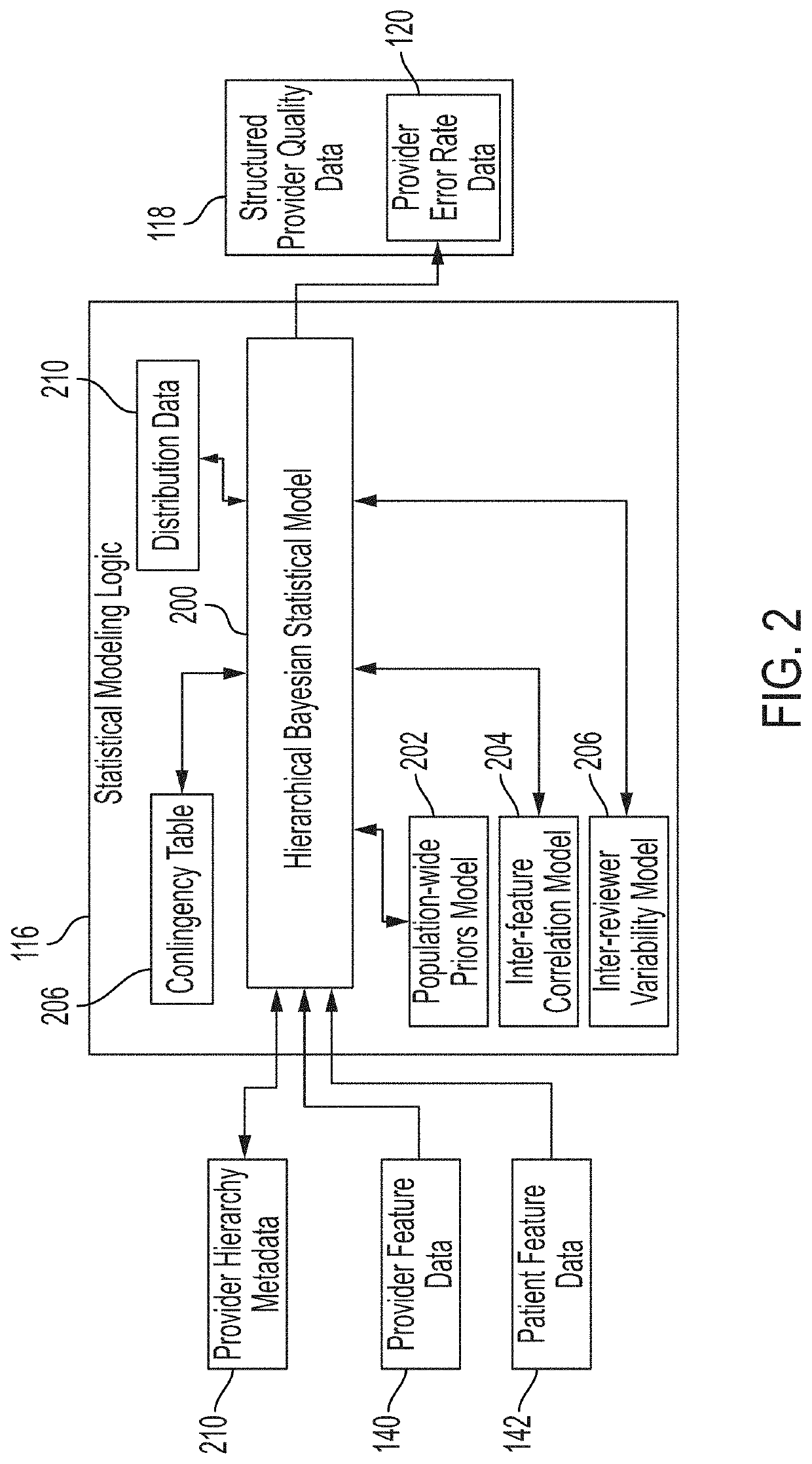
  • Computer-implemented machine learning for detection and statistical analysis of errors by healthcare providers
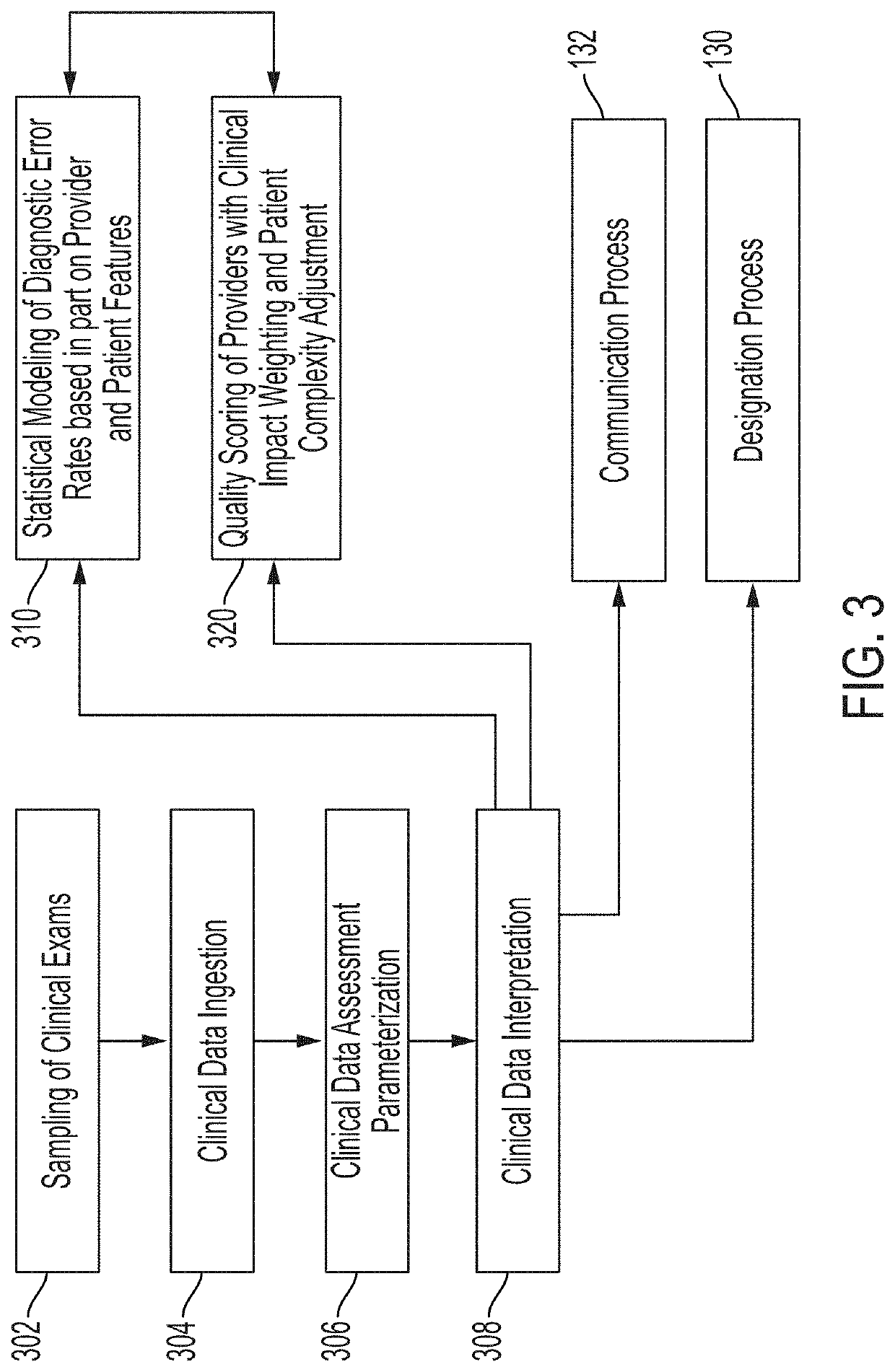
  • Computer-implemented machine learning for detection and statistical analysis of errors by healthcare providers

Examples


Embodiment Construction

[0048]Various embodiments of the disclosure are discussed in detail below. While specific implementations are discussed, it should be understood that this is done for illustration purposes only. A person skilled in the relevant art will recognize that other components and configurations may be used without parting from the spirit and scope of the disclosure. Additional features and advantages of the disclosure will be set forth in the description which follows, and in part will be obvious from the description, or can be learned by practice of the herein disclosed principles. It will be appreciated that for simplicity and clarity of illustration, where appropriate, reference numerals have been repeated among the different figures to indicate corresponding or analogous elements. The description is not to be considered as limiting the scope of the embodiments described herein.

[0049]Using various machine learning techniques and frameworks, it is possible to analyze data sets to extract ...


PUM


Abstract

For training data pairs comprising training text (a radiological report) and training images (radiological images associated with the radiological report), a first encoder network determines word embeddings for the training text. A concept is generated from the operation of layers of the first encoder network, which is regularized by a first loss between the generated concept and a labeled concept for the training text. A second encoder network determines features for the training image. A heatmap is generated from the operation of layers of the second encoder network, which is regularized by a second loss between the generated heatmap and a labeled heatmap for the training image. A categorical cross entropy loss is calculated between a diagnostic quality category (classified by an error encoder) and a labeled diagnostic quality category for the training data pair. A total loss function comprising the first, second, and categorical cross entropy losses is minimized.

Description

CROSS-REFERENCE TO RELATED APPLICATIONS[0001]This application claims the benefit of priority to U.S. patent application Ser. No. 16 / 386,006 filed Apr. 16, 2019 and entitled “COMPUTER-IMPLEMENTED DETECTION AND STATISTICAL ANALYSIS OF ERRORS BY HEALTHCARE PROVIDERS,” the disclosure of which is herein incorporated by reference in its entirety.TECHNICAL FIELD[0002]The present disclosure relates generally to computer-implemented machine learning systems and methods that are programmed to classify digital image data alone or in combination with unstructured text data, and more specifically pertains to machine learning systems and methods for diagnostic error detection.BACKGROUND[0003]The approaches described in this section are approaches that could be pursued, but not necessarily approaches that have been previously conceived or pursued. Therefore, unless otherwise indicated, it should not be assumed that any of the approaches described in this section qualify as prior art merely by virt...

Claims


Application Information

Patent Timeline
IPC IPC(8): G06T7/00G06K9/62G06N3/04G06N3/08G16H30/20
CPCG06T2207/20081G06K9/6256G06N3/0445G16H30/20G06N3/084G06T7/0012G06T2207/20084G06K9/6215G16H50/20G16H30/40G06V30/40G06V10/98G06V2201/03G06V10/82G06V10/803G06V30/1916G06V30/19173G06V30/19147G06N7/01G06N3/048G06N3/044G06N3/045G06F18/251G06V30/333G06V30/133G06F18/214G06F18/22
Inventor VIANU, RONAIJO, TARMO HENRIKBROWNING, JAMES ROBERTDONG, XIAOJINEAKIN, BRYCE ERONELGORT, DANIEL ROBERTHERZOG, RICHARD J.ODRY, BENJAMIN L.PARK, JINHYEONGSUUTARI, BENJAMIN SELLMANDUBBIN, GREGORY ALLEN
Owner COVERA HEALTH
Why Eureka
Eureka Blog
Learn More