Three-dimensional printed bone supported sinus guide for edentulous maxillary arch

a three-dimensional printing and bone supported technology, applied in the field of surgical guides, can solve the problems of inability of the operator to precisely locate the floor of the sinus, lack of bony support around the portion of the implant, and frustrate the purpose of stably anchoring dental implants, etc., and achieve the effect of accurately preparing an osteotomy

Active Publication Date: 2020-10-08
IMAM ABDULRAHRNAN BIN FAISAL UNIVERSITY
View PDF0 Cites 0 Cited by
  • Summary
  • Abstract
  • Description
  • Claims
  • Application Information

AI Technical Summary

Benefits of technology

The invention is a surgical guide for sinus elevation procedures that is custom shaped and dimensioned based on a CT scan and 3-D imaging software results. The guide allows a surgeon to accurately prepare an osteotomy and access the sinus cavity without damage to the underlying membrane. The guide eliminates the need for approximation of the osteotomy, ensuring easy access to the sinus cavity. The guide can be produced based on the position of a tooth implant or a window covering the sinus. It allows for smooth contouring of the surgical guide and can be further improved in its design.

Problems solved by technology

This results in a lack of bony support around that portion of the implant and frustrates the purpose of stably anchoring dental implants in the maxilla.
However, historically, Caldwell-Luc technique and other techniques for sinus approaches did not mention guides to be used during a surgical procedure.
One of the technical difficulties encountered during this procedure is the inability of the operator to precisely locate the floor of the sinus while preparing the osteotomy from an antero-posterior direction (along the X-Y axis).
Since the floor of the sinus can elevate and descend variably as the osteotomy moves antero-posteriorly, it is impossible to visualize this course.
The additional trimming of bone is traumatic and unnecessarily removes bone.
Another error occurs if the osteotomy is placed too superior to the floor of the sinus.
This technically difficult maneuvering of the instruments along two planes increases the risk of tearing the membrane and thus compromising the outcome of the graft.
This would lead to additional trimming of bone and increase the risk of tearing the membrane during the expansion of the osteotomy.
It is nearly impossible to visualize the variable course of the sinus floor as the osteotomy progresses antero-posteriorly.
This inability to visualize the course of the sinus floor is the first difficulty encountered in the procedure.
Another difficulty encountered is the varying thickness of the lateral wall of the sinus as the osteotomy penetrates it to expose the underlying Schneiderian membrane.
However, if the osteotomy is prepared too deep, it can tear through the fragile membrane.
A further difficulty encountered is the anterior wall of the sinus.
This would require manipulation anteriorly and then laterally to dissect the membrane from the lateral and anterior walls thus increasing the risk of tearing the membrane from the difficult manipulation in two planes.
Again, any additional adjustments to the osteotomy would cause unnecessary bone removal and trauma as well as increase the risk of tearing the membrane.
During an actual osteotomy, it is nearly impossible to accurately follow the varying course of the sinus.
Inevitably there are areas that are not exposed by the osteotomy which require careful manipulation of the sinus membrane and risk damage to the membrane.
Nevertheless, even with the information provided by the CT scan utilizing the 3-D imaging software as to the outline of the sinus in the X, Y and Z planes and all other parameters, there has been no mechanism to accurately transfer this highly precise information to the surgical field for its practical application.
Meticulous planning of the parameters of the sinus to be elevated and augmented has been thwarted by the inaccurate approximation in the transfer of this information during the surgical procedure as well as inexact fabrication of surgical guides designed using this information.
Existing procedures proposed for making surgical guides, such as those described by Massoud, US 2010 / 0191242, can lack precision in the ways they produce a surgical guide, for example, the surgical guides are produced by methods that involve use of plastics that require heat curing such as heat-cure acrylic resin or which cause shrinkage in a final produced surgical guide and thus introduce inaccuracy into the guide compared to the extremely precise imaging information used to design the guide.
These deformations and distortions of the original imaging information affect the accuracy of a surgical procedure using the guide.
Due to the anatomical features of the maxilla and surrounding sites, even small distortions in a conventional surgical guide can produce rocking and ill-fitting guides which lack accuracy and can dramatically affect the X, Y and Z locations identifying a surgical procedure site, such as sites for sinus window preparation sites for dental implants.
This lack of accuracy can displace the desired site of surgery identified by a medical scan to an inappropriate site having insufficient grafting materials or support.
Thus such distortions can lead to the need for additional corrective surgery.
Moreover, the lack of surgical guide precision can also cause irreversible injury to vital structures such as the maxillary artery or inferior alveolar nerve and increase patient discomfort and pain by irritating the adjacent teeth and gums.
Often these patients experience significant resorption of facial bone along with loss of teeth.
These patients already have an increased risk of insufficient bone material to support implants and can require more intensive surgical grafting procedures.
Poor bone quality in these patients as well as hidden features such as sinus cavities, nerves and nerve roots complicate dental implant procedures.
A variety of different UV-curable resins is known, however, a primary limitation of their use is the limited light penetration depth which usually does not exceed a few millimeters.
However, these are subject to oxygen inhibition where oxygen in air can stop surface molecules from polymerizing leaving a tacky layer.
However, only a limited number of monomers and oligomers are available for light induced cationic cure.
Thus, the versatility in tailoring properties is rather limited.
Furthermore, the photoinitiators used in cationic cure can be toxic and corrosive and cationic systems not subjected to air inhibition are easily poisoned by moisture in the air.

Method used


Image

  • Three-dimensional printed bone supported sinus guide for edentulous maxillary arch
  • Three-dimensional printed bone supported sinus guide for edentulous maxillary arch
  • Three-dimensional printed bone supported sinus guide for edentulous maxillary arch

Examples


example

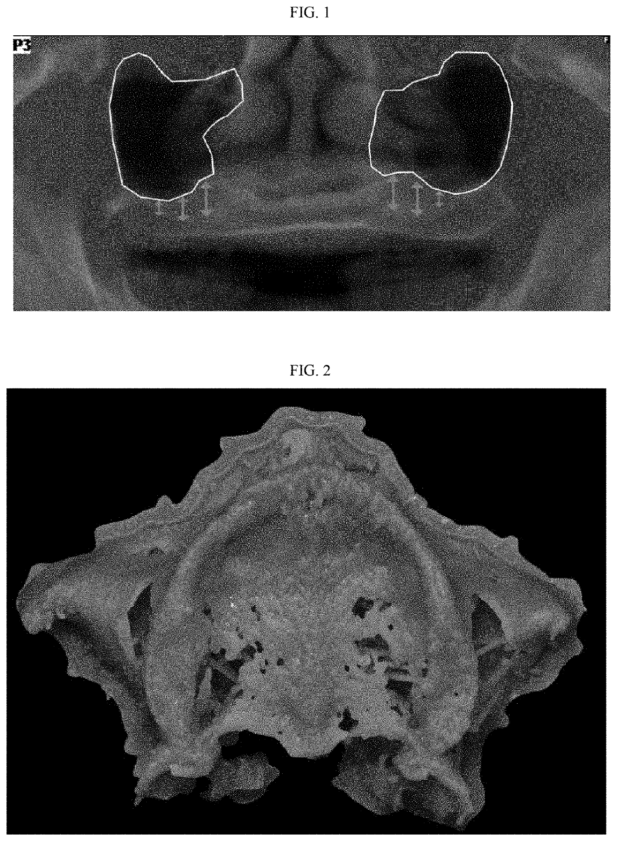
[0084]A CBCT scan of a patient's maxilla and jaw is taken using a VGi Evo instrument (https: / / _www.cefladental.com / our-brands / newtom / , last accessed Mar. 5, 2019, incorporated by reference) to diagnose and plan treatment of an edentulous patient. The CBCT cross section is used to evaluate and plan sinus grafting to support a dental implant. Data from the scan is provided in a DICOM file which can be used to provide a 3D model of the maxillary bone.

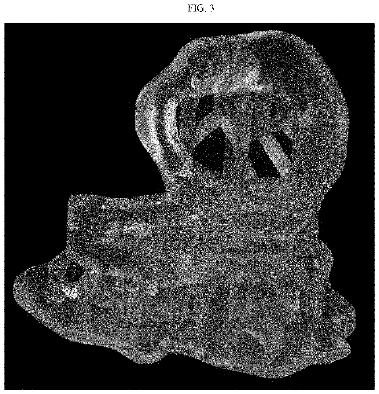
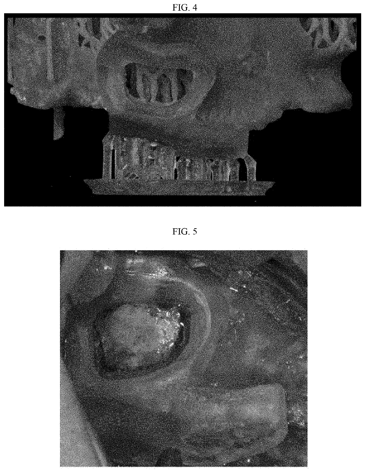
[0085]DICOM reconstruction software (InVesalius 3.0) is used to transform that DICOM data set from the CBCT scan into a 3D bone model. The bone model is exported as an STL file.

[0086]The STL file depicting maxillary bone is superimposed on the CBCT data using implant-planning software (BlueSky Plan; Imps: / / _blueskybio.com / pages / blue-sky-plan-guided-surgery-software, last accessed Mar. 5, 2019, incorporated by reference). Implant position is selected and a gum-bone supported surgical guide is designed which includes the implant position and...


PUM


Abstract

An accurate and precise surgical guide for dental procedures, especially a procedure involving a subject with few or no teeth as well as methods for making and use of such guides.

Description

BACKGROUND OF THE INVENTIONField of the Invention[0001]The invention relates to a surgical guide for use in the fields of dental surgery and prosthetics. More specifically the invention relates to a surgical guide for edentulous patients to be used in maxillary surgical procedures such as sinus elevation or grafting in preparation for installation of dental or endosseous implants which form intimate bonds to bone. These implants in turn support dental prosthesis such as a crown, bridge, denture, facial prosthesis or act as orthodontic anchors.Description of Related Art[0002]The “background” description provided herein is for the purpose of generally presenting the context of the disclosure. Work of the presently named inventor to the extent it is described in this background section as well as aspects of the description which may not otherwise qualify as prior art at the time of filing, are neither expressly or impliedly admitted as prior art against the present invention.[0003]Foll...

Claims


Application Information

Patent Timeline
IPC IPC(8): A61C8/00A61B17/17A61B17/16
CPCA61C8/0092A61F2/186A61B17/1688A61B17/1615A61B17/1785A61B17/176A61C1/084A61F2/4644A61B34/10A61B2017/568A61C1/082A61B2034/105B33Y50/00B33Y80/00A61B17/24A61B2017/00526A61B2017/00743A61B2017/00831A61B2034/108
Inventor ALRUMAIH, HAMAD SALEH HAMAD
Owner IMAM ABDULRAHRNAN BIN FAISAL UNIVERSITY
Why Eureka
Try Eureka