Eye disease biomarker
a biomarker and eye disease technology, applied in the field of eye disease biomarkers, can solve the problems of difficult to identify a substance that causes inflammation, has not been identified, and cannot be directly identified, so as to achieve accurate evaluation of central serous chorioretinopathy, quantitatively evaluating corneal or conjunctival disease more conveniently, and accurate diagnosis
- Summary
- Abstract
- Description
- Claims
- Application Information
AI Technical Summary
Benefits of technology
Problems solved by technology
Method used
Image
Examples
experimental example 1
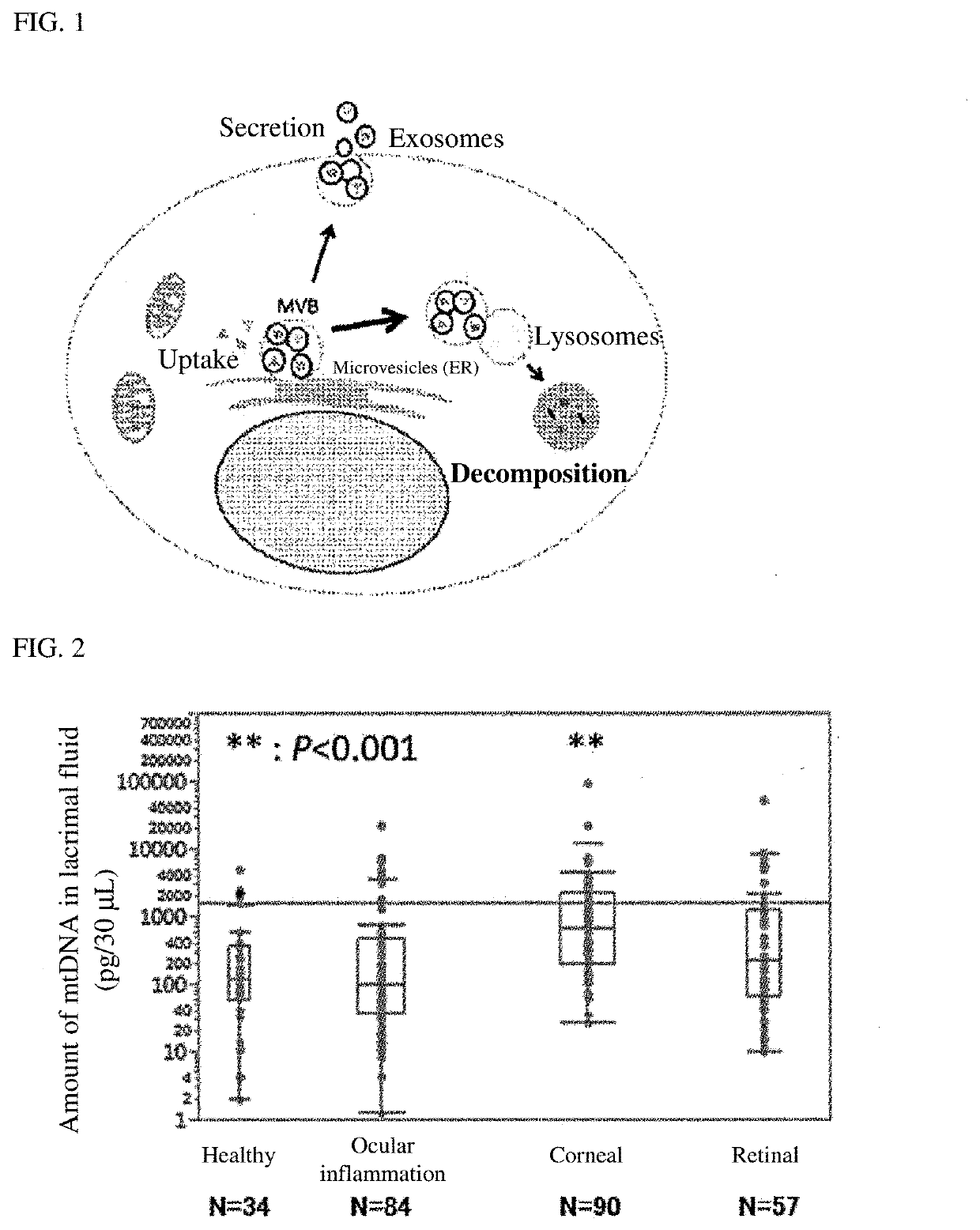
[0089]A patients having ocular inflammation (uveitis), corneal disease or retinal disease (age-related macular degeneration) was used as a subject. The amount of mitochondrial DNA (mtDNA) in lacrimal fluid of the subject was measured and evaluated in accordance with the following method. The amount of mtDNA in lacrimal fluid of each of healthy controls as a group of normal controls was measured under the same conditions. The subjects included 84 ocular inflammation patients, 90 corneal disease patients, 57 retinal disease patients and 34 normal persons (healthy controls).
Preparation of Lacrimal Fluid Sample
[0090]As a lacrimal fluid sample, 30 μL of PBS (phosphate buffer solution) used as a wiping liquid for the lower palpebral conjunctiva, and recovered was used. Specifically, 30 μL of PBS was taken with Pipetman, and a subject was made to turn up. While an observation was made with a slit-lamp microscope, the PBS was dropped onto the lower palpebral conjunctiva of the subject, and ...
experimental example 2
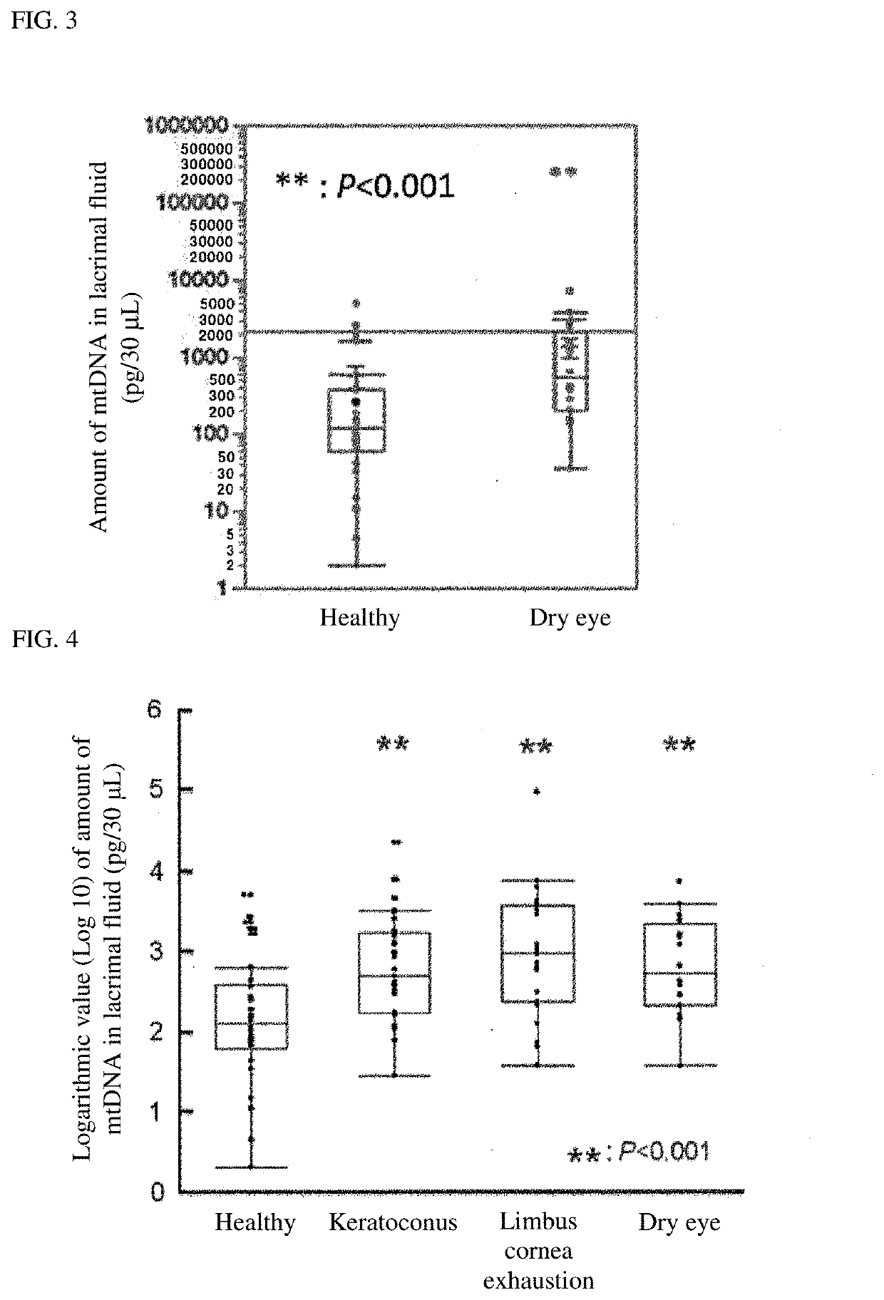
[0097]The amount of mtDNA in lacrimal fluid of each of dry eye patients (10 patients) was evaluated using the same method as in Example 1. The amount of mtDNA in lacrimal fluid of each of healthy controls (20 persons) as a group of normal controls was measured under the same conditions. The obtained mtDNA level was statistically analyzed by the Student's t test method. The results are shown in FIG. 3.
[0098]FIG. 3 is a graph showing mtDNA levels in lacrimal fluid of a group of dry eye patients and a group of normal controls. The graph in FIG. 3 shows that the mtDNA level in lacrimal fluid in the group of dry eye patients is significantly higher than that in the group of normal controls (P<0.001). This indicates that a dry eye patient has an increased mtDNA level in lacrimal fluid as compared to a group of normal persons (healthy controls), and thus mtDNA in lacrimal fluid can serve as a biomarker for dry eye.
experimental example 3
[0099]The expression level of mtDNA in lacrimal fluid of a subject with keratoconus, limbal corneal stem cell deficiency or dry eye was measured and evaluated by the same method as in Example 1. The expression level of mtDNA in lacrimal fluid of each of healthy controls as a group of normal controls was measured under the same conditions. The subjects included 39 keratoconus patients, 29 limbal corneal stem cell deficiency patients, 20 dry eye patients and 34 healthy controls. The expression level of mtDNA was statistically examined by the Steel test method after a relative value obtained based on a calibration curve was logarithmically processed. The results are shown in FIG. 4.
[0100]FIG. 4 is a graph showing expression levels of mtDNA in lacrimal fluid of a group of patients with keratoconus, limbal corneal stem cell deficiency or dry eyes and a group of normal controls. The graph in FIG. 4 shows that the expression level of mtDNA in lacrimal fluid in each of the groups of patient...
PUM
| Property | Measurement | Unit |
|---|---|---|
| Volume | aaaaa | aaaaa |
Abstract
Description
Claims
Application Information
 - R&D
- Intellectual Property
- Life Sciences
- Materials
- Tech Scout
- Unparalleled Data Quality
- Higher Quality Content
- 60% Fewer Hallucinations
Browse by: Latest US Patents, China's latest patents, Technical Efficacy Thesaurus, Application Domain, Technology Topic, Popular Technical Reports.
© 2025 PatSnap. All rights reserved.Legal|Privacy policy|Modern Slavery Act Transparency Statement|Sitemap|About US| Contact US: help@patsnap.com


