
Graphene-based electro-microfluidic devices and methods for protein structural analysis

a technology of microfluidic devices and graphene, which is applied in the field of microfluidic devices and xray analysis, can solve the problems of large-scale serial methods, large number of high-quality crystals to grow and manipulate, and problems that are then further compounded, so as to achieve faster nucleation and crystal growth, and higher signal-to-noise

Active Publication Date: 2019-12-19
UNIV OF MASSACHUSETTS
View PDF0 Cites 2 Cited by
  • Summary
  • Abstract
  • Description
  • Claims
  • Application Information

AI Technical Summary

Benefits of technology

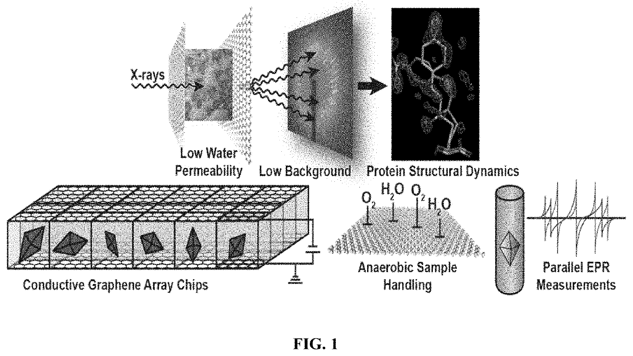
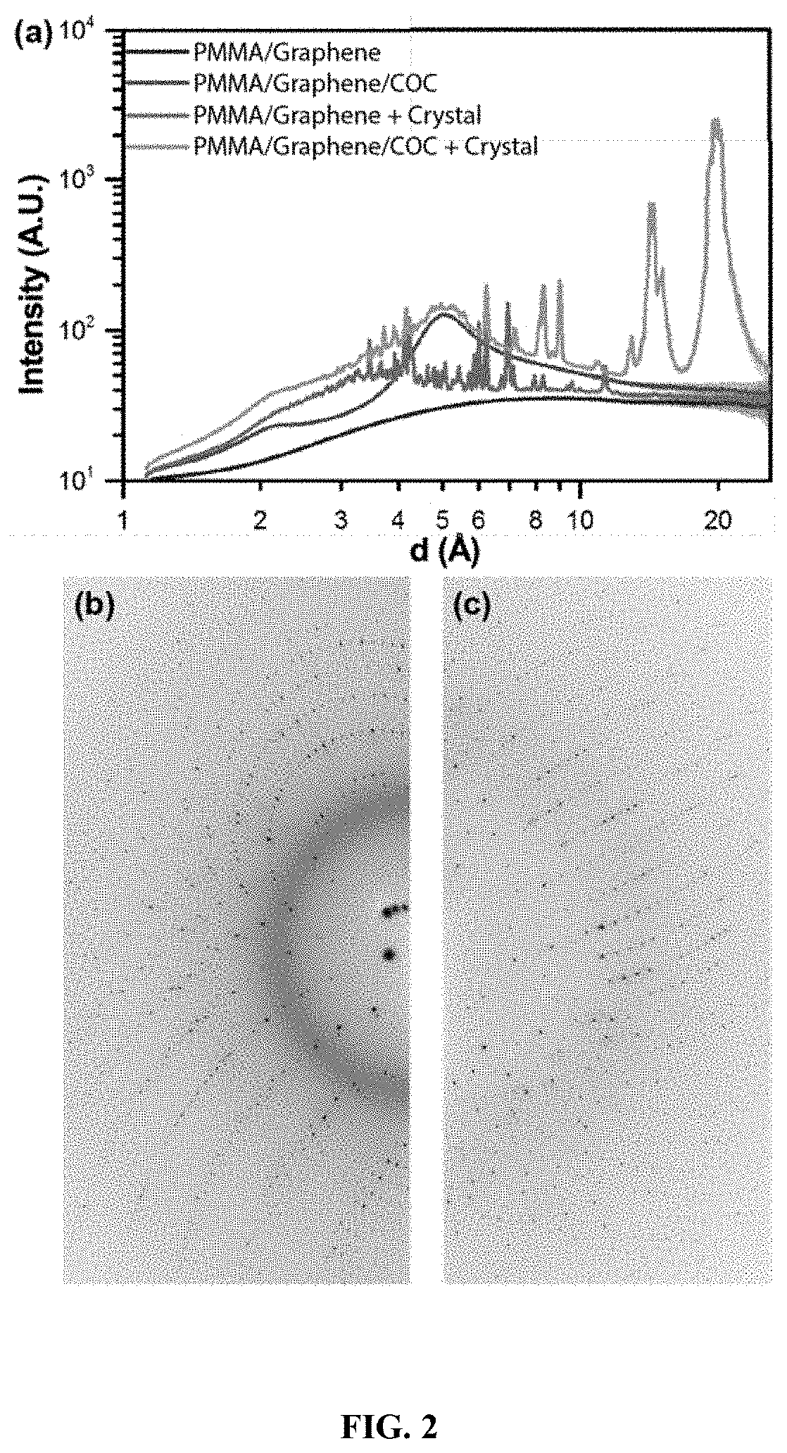
The patent describes a new device that uses ultra-thin films made of graphene to create and analyze crystals using a process called electro-crystallization. The device also includes an X-ray transparent window so that in situ X-ray analysis can be done while the crystals form. This method allows for faster sodiumation and crystal growth, as well as higher signal-to-noise for diffraction data obtained from crystals in the presence of an electric field. The device can also be used to examine the effect of an electric field on the structure and dynamics of the crystal.

Problems solved by technology

In the meantime, the challenges of such platforms lie in the need to maintain a protected sample environment as well as to minimize the interference of device materials with the subsequent X-ray analysis.
These largescale serial methods, however, suffer from the need to grow and manipulate a large number of high quality crystals.
These issues are then further compounded by the need to deliver such samples efficiently to the X-ray beam and the challenge of synchronizing structural dynamics within crystals.
Major challenges remain in microfluidic X-ray analysis and protein crystallography.

Method used


Image

  • Graphene-based electro-microfluidic devices and methods for protein structural analysis
  • Graphene-based electro-microfluidic devices and methods for protein structural analysis
  • Graphene-based electro-microfluidic devices and methods for protein structural analysis

Examples


examples

Graphene Film Preparation

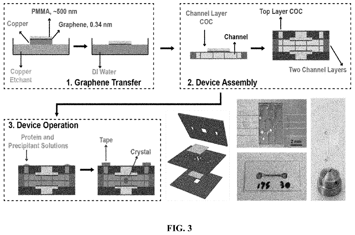
[0080]Large area graphene was synthesized on a copper substrate (Graphene Platform) by chemical vapor deposition in a quartz tube furnace (Planar Tech) using standard methods [48-51]. After synthesis, the back side of the copper substrate was scrubbed with a Kimwipe to remove residual graphene. Patterning of the graphene electrodes was achieved using two different methods. The first method simply used thin-tip tweezers (TDI International Inc.) to scratch a narrow line into the graphene / copper film. The second method defined the desired structure of the electrodes using a protective mask made from a piece of thermal release tape (Semiconductor Equipment Corp.) cut to the desired shape using a cutting plotter (Graphtech CE6000), followed by a 5-minute etching of the exposed graphene by an oxygen plasma (Harrick Plasma). Following patterning of the graphene electrodes, a roughly 500-nm thick layer of poly(methylmethacrylate) (950PMMA A4, Microchem) was then spi...


PUM

PropertyMeasurementUnit
Thicknessaaaaaaaaaa
Thicknessaaaaaaaaaa
Volumeaaaaaaaaaa

Abstract

The invention provides a novel microfluidic platform for use in electro-crystallization and electro-crystallography experiments. The manufacturing and use of graphene as X-ray compatible electrodes allows the application of electric fields on-chip, during X-ray analysis. The presence of such electric fields can be used to modulate the structure of protein (or other) molecules in crystalline (for X-ray diffraction) or solution form (for X-ray scattering). Additionally, the presence of an electric field can be used to extend the lifetime of fragile samples by expediting the removal of reactive secondary radiation damage species.

Description

PRIORITY CLAIMS AND RELATED PATENT APPLICATIONS[0001]This application claims the benefit of priority to U.S. Provisional Application Ser. No. 62 / 687,190, filed on Jun. 19, 2018, the entire content of which is incorporated herein by reference in its entirety.STATEMENT REGARDING FEDERALLY FUNDED RESEARCH OR DEVELOPMENT[0002]This invention was made with government support under Grant No. NSF-1231306 awarded by the National Science Foundation. The Government has certain rights in the invention.TECHNICAL FIELD OF THE INVENTION[0003]The invention generally relates to microfluidic devices and X-ray analysis. More particularly, the invention relates to electro-microfluidic devices that utilize graphene films as architectural materials and methods of fabrication and use thereof in X-ray analysis of target materials (e.g., biomolecules).BACKGROUND OF THE INVENTION[0004]Microfluidic and microscale devices have a demonstrated potential to enable both protein crystallization and in situ X-ray di...

Claims


Application Information

Patent Timeline
IPC IPC(8): G01N27/26B01L3/06B01L3/00C30B7/14G01N23/20025G01N23/207B01D9/00
CPCG01N27/26B01L3/06G01N23/20025B01L3/502761B01D9/0072B01L3/502707C30B7/14G01N23/207B01L2200/10B01D9/0063B01D9/0077B01L3/502715B01L2300/0645B01L2300/0887C30B7/12C30B29/58G01N23/20G01N23/20008G01N23/20066G01N23/20083G01N23/20091G01N23/201G01N23/203G01N23/205G01N23/2076G01N23/22G01N23/2202G01N23/2204G01N23/2206G01N23/2208G01N23/223G01N23/2209
Inventor PERRY, SARAH L.SUI, SHOU
Owner UNIV OF MASSACHUSETTS
Learn More