The application of anaplasma phagocytophilum protein aph1384
a technology of anaplasma phagocytophilum and anaplasma phagocytophilum, which is applied in the field of immunology, can solve the problems of high cost of antigen slides, epidemic levels of granulocytic anaplasmosis worldwide, and inability to detect granulocytic anaplasmosis in time, so as to facilitate rapid and accurate clinical diagnosis and improve detection sensitivity. , the effect of improving the sensitivity of detection
- Summary
- Abstract
- Description
- Claims
- Application Information
AI Technical Summary
Benefits of technology
Problems solved by technology
Method used
Image
Examples
embodiment 1
[0015]Preparation of APH1384 protein (a species-specific protein derived from Anaplasma phagocytophilum)
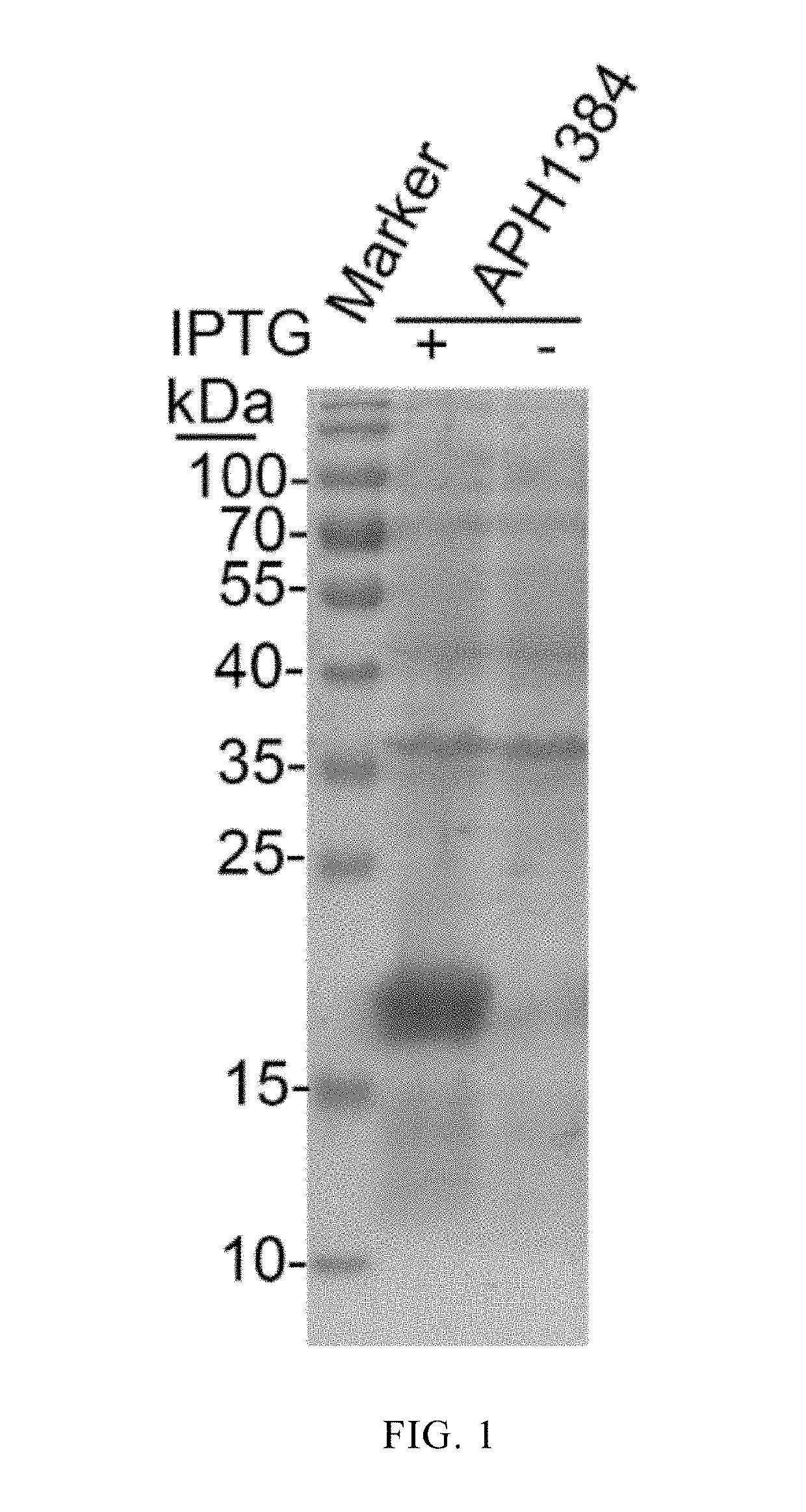
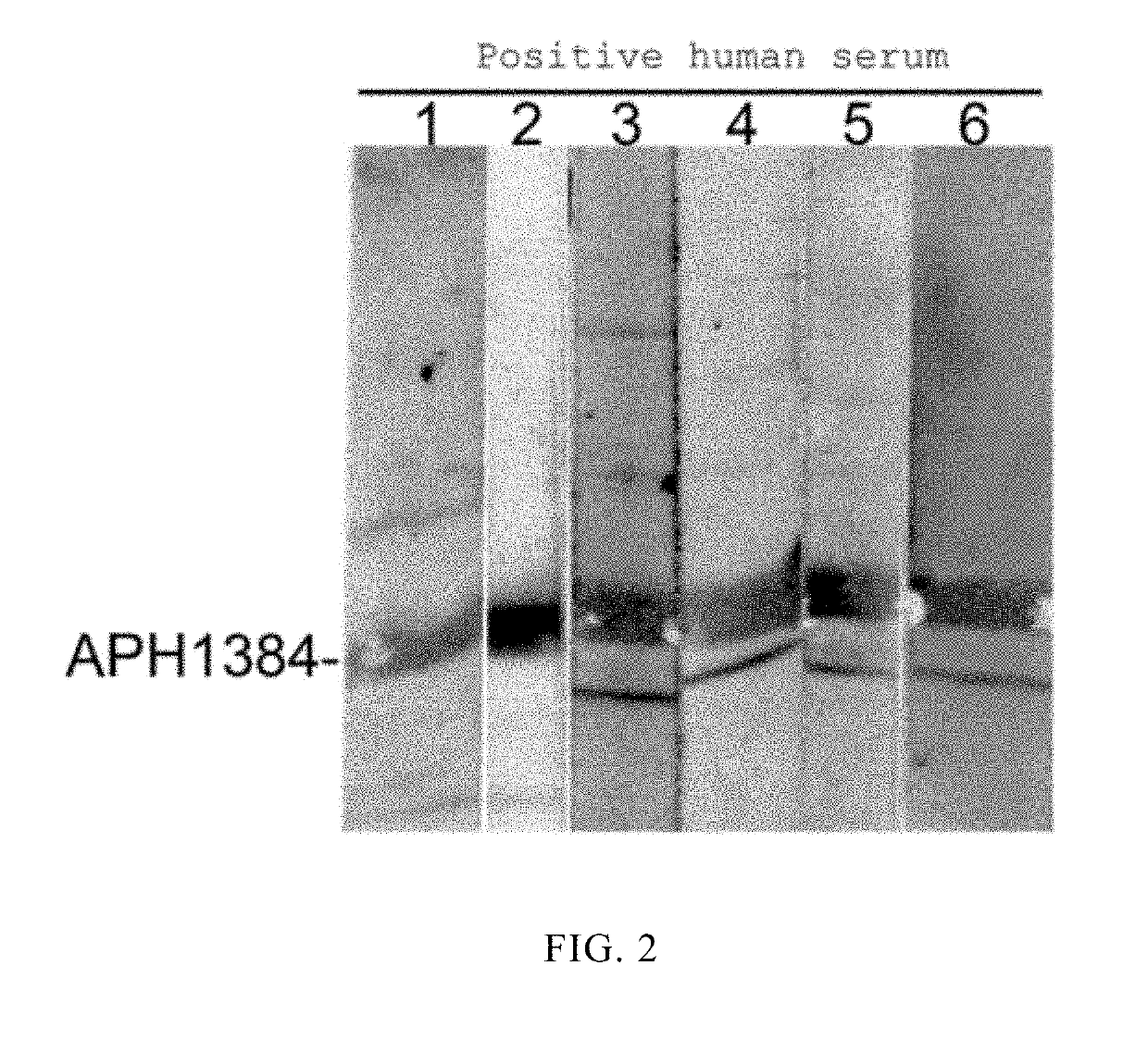
[0016]Species-specific proteins derived from Anaplasma phagocytophilum were screened by bioinformatics techniques using 610 proteins of unknown function encoded by the genome of Anaplasma phagocytophilum as targets. Some of the proteins (including APH1384) were subjected to cloning and expression, and antigenic identification. It was found from the research results that APH1384 had strong antigenicity. Detailed steps of cloning and expression, and antigenic characterization of APH1384 were provided as below. Specific primers containing restriction endonuclease sites were designed according to a gene sequence aph1384 in GenBank: aph1384-F-TCCGAATTCATGTTTGATATATTTAATGATGCTG; and aph1384-R-GCACTCGAGTTATTCGGACGCAGAGGCT. A DNA fragment of the gene aph1384 was amplificated by PCR using whole genomic DNA of Anaplasma phagocytophilum as a template. The amplificated target gene DNA fragmen...
embodiment 2
[0018]Preparation of APH1384 Polypeptide
[0019]An antigenic epitope of APH1384 was predicted and obtained by bioinformatics methods, and amino acid sequence of the antigenic epitope is shown in SEQ ID NO: 2. An APH1384 antigenic epitope polypeptide was chemically synthesized, and antigenicity of the APH1384 polypeptide was verified by DB technique. Specific operations were provided as below.
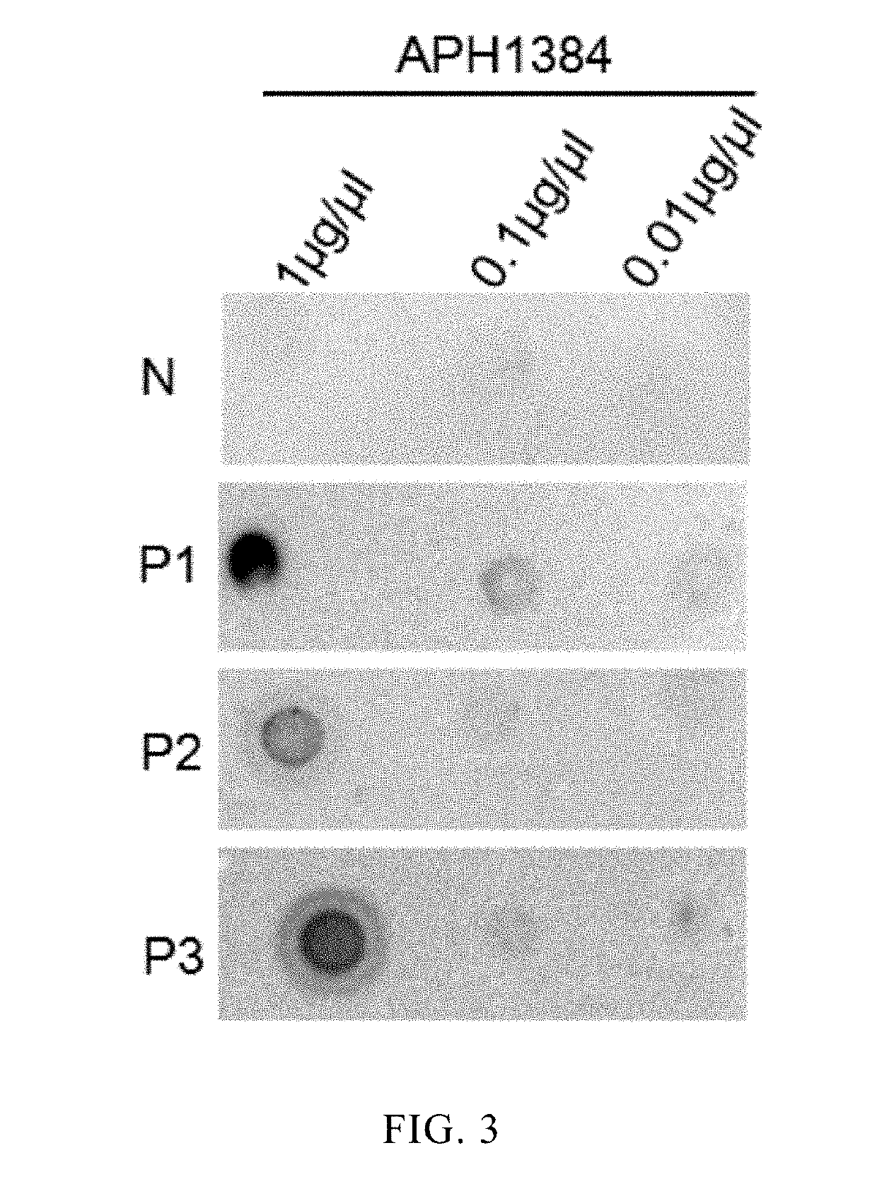
[0020]Dilutions of the APH1384 polypeptide antigen (1 μl at concentrations of 1 μg / l, 0.1 μg / l, and 0.01 μg / μl respectively) were added to a nitrocellulose membrane respectively, and the membrane was dried for 20-30 min at 37° C., blocked for 30 min, incubated with primary human positive sera (1:2000) for 1 h, incubated with a secondary antibody (1:5000) for 1 h, and developed. The results are shown in FIG. 3, wherein N represents a negative serum, and P1-P3 each represents one Anaplasma phagocytophilum positive serum. The results show that the negative serum does not react with APH1384, and three...
embodiment 3
[0021]Use of APH1384 Polypeptide
[0022]Specific anti-APH1384 antibodies in human sera were detected by ELISA with the synthesized APH1384 polypeptide as coating antigen. Specific operation steps were provided as below:
[0023](1) coating: the APH1384 polypeptide synthesized in example 2 was diluted to 20 μg / ml with PBS, and coated onto a plate overnight at 4° C. or for 2 h at room temperature (96-well plate, 50 μl / well);
[0024](2) blocking: the 96-well plate was washed 3 times with PBS, and a blocking solution of bovine serum albumin (at a concentration of 1% (w / v)) was added in 200 μl / well at 37° C. for 2 h;
[0025](3) incubation with a primary antibody: the serum to be detected was diluted at a ratio of 1:1000 using the blocking solution, and the dilution was added in 100 μl / well and incubated for 1 h at 37° C.;
[0026](4) incubation with a secondary antibody: the 96-well plate was washed 4 times with PBS (200 μl / well), and the secondary antibody (HRP labeled goat anti-human IgG, 1:5000 d...
PUM
| Property | Measurement | Unit |
|---|---|---|
| Sensitivity | aaaaa | aaaaa |
Abstract
Description
Claims
Application Information
 - R&D
- Intellectual Property
- Life Sciences
- Materials
- Tech Scout
- Unparalleled Data Quality
- Higher Quality Content
- 60% Fewer Hallucinations
Browse by: Latest US Patents, China's latest patents, Technical Efficacy Thesaurus, Application Domain, Technology Topic, Popular Technical Reports.
© 2025 PatSnap. All rights reserved.Legal|Privacy policy|Modern Slavery Act Transparency Statement|Sitemap|About US| Contact US: help@patsnap.com


