
Enhanced gene delivery methods

a technology of gene delivery and gene-correction, applied in the field of enhanced gene delivery methods, can solve the problems of cell death, inefficient gene-correction attempts in these and other cells, etc., and achieve the effect of reducing the transfection or transfection efficiency and targeting cell transduction or transfection efficiency

Pending Publication Date: 2019-05-02
ANGIOCRINE BIOSCI
View PDF0 Cites 0 Cited by
  • Summary
  • Abstract
  • Description
  • Claims
  • Application Information

AI Technical Summary

Benefits of technology

The patent text describes a method of improving the efficiency of gene delivery to target cells, such as stem cells, by using endothelial cells that have been optimized to be cultured ex vivo. The method results in a similar level of target cell transduction or transfection efficiency even when compared to a control group without the endothelial cell co-culture. This can be achieved even when the amount of exogenous nucleic acid molecules supplied to the target cells is the same or reduced because of the presence of the co-cultured endothelial cells. Overall, this method provides a reliable and effective way to deliver genes to target cells.

Problems solved by technology

Unfortunately, attempts at gene-correction in these and other cells have thus far proven to be largely inefficient.
Several cell transduction and transfection techniques (such as electroporation) cause significant cell stress and cell damage, often resulting in cell death.
This is a particular problem for gene delivery to stem cells, and is also a particular problem in applications that require large quantities of healthy, viable, transduced or transfected cells.

Method used


Image

  • Enhanced gene delivery methods
  • Enhanced gene delivery methods
  • Enhanced gene delivery methods

Examples


example 1

Effect of E4ORF1+Co-Culture on Expansion & Viral Transduction of HSPCs

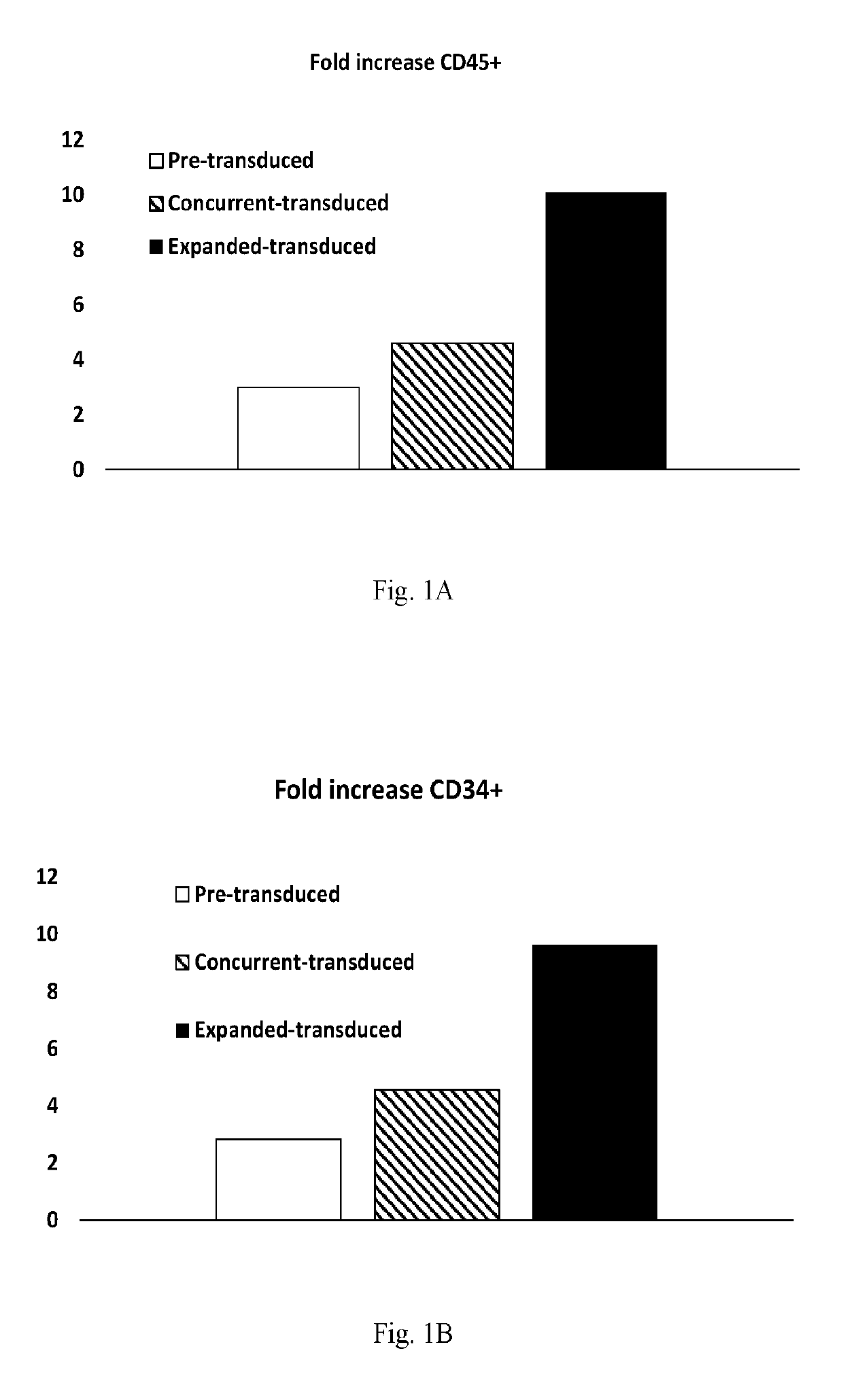
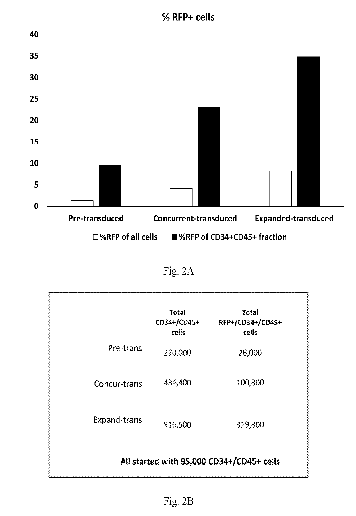
[0080]Experiments were performed to assess the effects of E4ORF1+ endothelial cells on the ability to expand and transduce CD34+ / CD45+ HSPCs. CD34+ cells (Lonza) were transduced with Red Fluorescent Protein (RFP) and expanded on E4ORF1+ umbilical vein endothelial cell (UVEC) cultures in 6-well plates using the three different protocols, as follows:

[0081]In protocol 1, transduction was commenced 24 hrs prior to starting HSPC-E4ORF1+ UVEC co-culture (“Pre-transduced”).

[0082]In protocol 2, transduction was performed concurrently with HSPC-E4ORF1+ UVEC co-culture (“Concurrent-transduced”).

[0083]In protocol 3 HSPCs were co-cultured with E4ORF1+ UVEC for 48 hrs, after which floating HSPCs were transferred to an empty well for transduction (24 hrs), and then following transduction the HSPCs were co-cultured with E4ORF1+ UVEC (“Expanded-transduced”).

In each of the above protocols cell culture was performed under hypoxic c...

example 2

Effect of Floating Fraction & Cytokines on Viral Transduction Efficiency

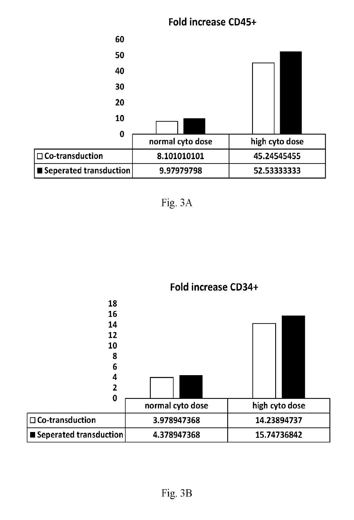
[0085]Experiments were performed to determine the effect of removing the ‘floating fraction’ of CD34+ hematopoietic cells on transduction efficiency and to determine if cytokine concentration has an influence on transduction efficiency.

[0086]CD34+ cells (Lonza) were transduced with Blue Fluorescent Protein (BFP) and expanded on E4ORF1+ UVEC cultures in 6-well plates using the following 4 different protocols, as follows:

[0087]In protocols 1 and 3, HSPCs were expanded on E4ORF1+ UVEC cultures for 96 hours and then subsequently transduced by incubating the HSPCS with lentivirus engineered to express BFP for 24 hours—while the HSPCs were still on the E4ORF1+EC feeder layer. The HSPCs were then expanded on the E4ORF1+ UVEC cultures for a further two days. Results obtained using these protocols are referred to in the figures as “co-transduction” results

[0088]In protocols 2 and 4, HSPCs were expanded on E4ORF1+ UVEC cu...

example 3

Rescue of Transfected Cells by Subsequent EC Co-Culture

[0093]Transfected or transduced cells, such as cells transfected by electroporation, can be cultured with ECs after transfection or transduction. Such subsequent co-culture can “rescue” cells that have been damaged by the transfection or transduction process—reducing cell death and loss of the transfected or transduced cells. Such co-culturing should be commenced “immediately” after the transfection or transduction step is commenced (as that term is defined in this patent specification), and should continue for sufficient time to allow recovery of the target cells. Without wishing to be bound by theory, it is believed that such “rescue” can occur as a result of cell-to-cell contact between the two cell populations (i.e. the transduced / transfected cells and the ECs), and / or as a result of exposure of the transduced / transfected cells to soluble factors secreted by the ECs, such as angiocrine factors and cytokines. The co-culture o...


PUM

PropertyMeasurementUnit
Fractionaaaaaaaaaa
Fractionaaaaaaaaaa
Defectsaaaaaaaaaa

Abstract

The present invention provides improved methods for gene delivery to, or genetic modification of target cells, wherein the gene delivery or other genetic modification of the target cells is performed in the presence of endothelial cells, or after co-culture of the target cells with endothelial cells, or wherein co-culture of the target cells with endothelial cells is employed immediately alter gene delivery in order to “rescue” cells that may have been damaged during the gene delivery process. In some embodiments gene delivery is performed by transfection. In some embodiments gene delivery is performed by transduction, in some embodiments the endothelial cells are organ-specific endothelial cells. In some embodiments the endothelial cells are E40RF1-expressing endothelial cells (E40RF1+ ECs). In some embodiments the target cells are stem cells, such as hematopoietic stem cells.

Description

CROSS-REFERENCE TO RELATED APPLICATIONS[0001]This application claims the benefit of priority of U.S. Provisional Patent Application No. 62 / 323,476 filed on Apr. 15, 2016, and U.S. Provisional Patent Application No. 62 / 403,110 filed on Oct. 1, 2016, the contents of each of which are hereby incorporated by reference in their entireties.INCORPORATION BY REFERENCE[0002]For the purpose of only those jurisdictions that permit incorporation by reference, all of the references cited in this disclosure (including but not limited to publications, patent applications, patents, and other references) are hereby incorporated by reference in their entireties. In addition, any manufacturers' instructions or catalogues for any products cited or mentioned herein are incorporated by reference. Many of the teachings provided in U.S. Pat. No. 8,465,732 can be used in conjunction with the present invention, or can be adapted for use with the present invention. Accordingly, the entire contents of U.S. Pat...

Claims


Application Information

Patent Timeline
IPC IPC(8): C12N15/87C12N15/86A61K35/545
CPCC12N15/87C12N15/86A61K35/545A61K35/28A61K35/44C12N2740/16043A61K35/12C12N5/10
Inventor FINNEGAN, PAUL WILLIAMDAVIS, CLAUDE GEOFFREYGINSBERG, MICHAEL DANIELNOLAN, DANIEL JOSEPH
Owner ANGIOCRINE BIOSCI
Learn More