System and method for local three dimensional volume reconstruction using a standard fluoroscope

a fluoroscope and volume reconstruction technology, applied in the field of system, apparatus, and method of navigation, position confirmation and position correction for surgical procedures, can solve the problems of difficult resolving of small soft tissue objects of interest such as lesions in fluoroscopic images, high cost of mri system or ct-based imaging system, and tedious procedures, so as to improve the quality of three dimensional reconstruction, increase the accuracy of camera pose, and increase the effect of accuracy

Active Publication Date: 2018-06-14
TYCO HEALTHCARE GRP LP
View PDF4 Cites 6 Cited by
  • Summary
  • Abstract
  • Description
  • Claims
  • Application Information

AI Technical Summary

Benefits of technology

The patent describes a system and method for creating three dimensional data of small soft-tissue objects in a video stream from a standard fluoroscopic imaging device. This data can be used for guidance, navigation planning, and treatment confirmation. The method involves creating a three-dimensional pseudo-volume by filtering out the obscuring objects based on their position and then projecting them back into two dimensions. The system uses multiple fluoroscopic images captured from different angles to improve the accuracy of the data. Additionally, the method compensates for patient body movement or jittering during rotation by using a single marker or catheter tip to stabilize the camera poses.

Problems solved by technology

An MRI system or CT-based imaging system is extremely costly, and in many cases not available in the same location as the location where a navigation plan is generated or where a navigation procedure is carried out.
However, although standard fluoroscopic images display highly dense objects such as metal tools and bones as well as large soft-tissue objects such as the heart, the fluoroscopic images have difficulty resolving small soft-tissue objects of interest such as lesions.
This is a tedious procedure as it requires several full CT scans, a dedicated CT room and blind navigation between scans.
Another option is a Cone-beam CT machine which is available in some operation rooms and is somewhat easier to operate, but is expensive and like the CT only provides blind navigation between scans, requires multiple iterations for navigation and requires the staff to leave the room.
The angle range may be very small (˜30°) which may result in poor three dimensional reconstruction quality.
Unfortunately, due to filtering, the angle measurement device may still report smooth angles throughout the video, ignoring the high-frequencies which are present in the actual camera poses.

Method used


Image

  • System and method for local three dimensional volume reconstruction using a standard fluoroscope
  • System and method for local three dimensional volume reconstruction using a standard fluoroscope
  • System and method for local three dimensional volume reconstruction using a standard fluoroscope

Examples


Embodiment Construction

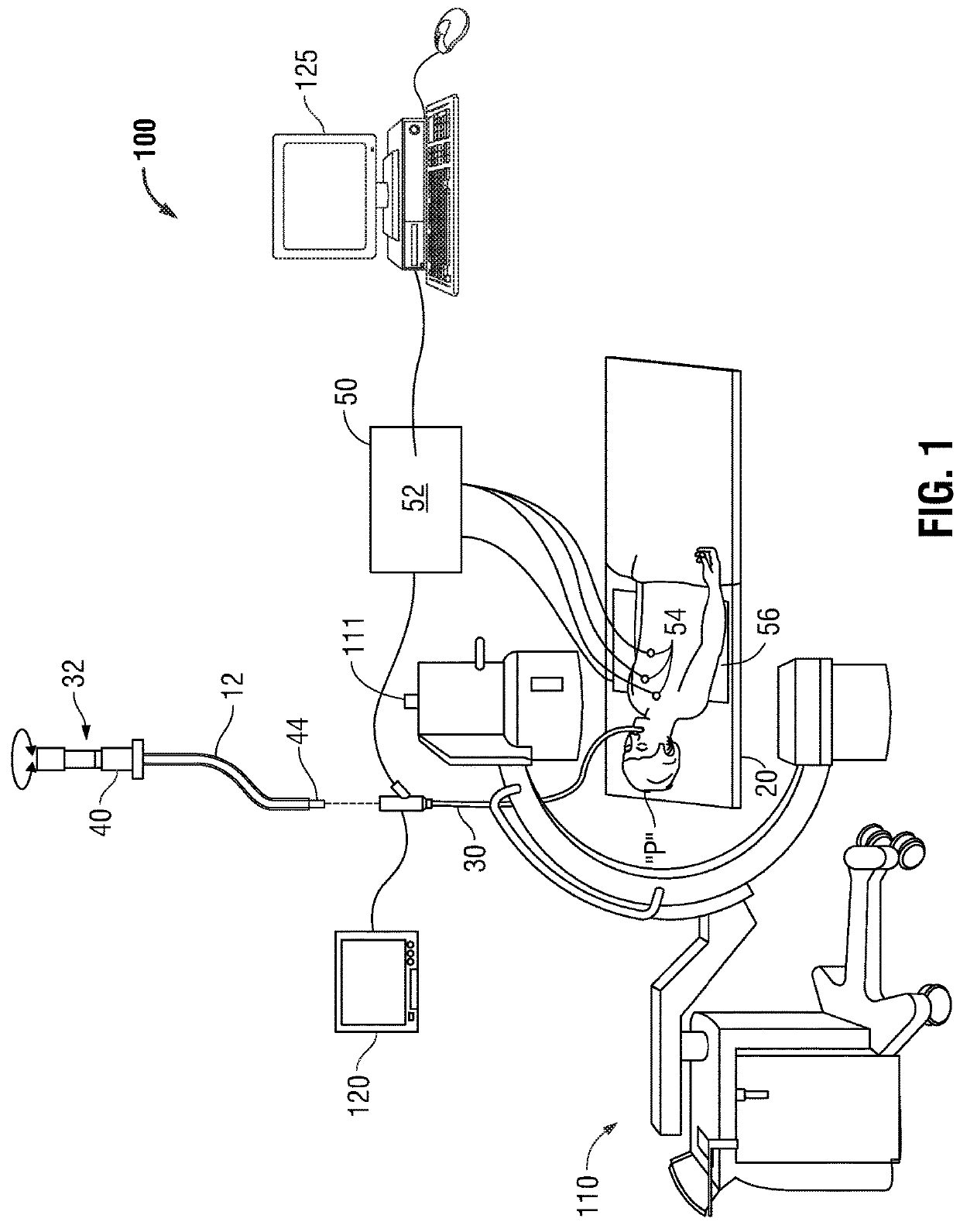
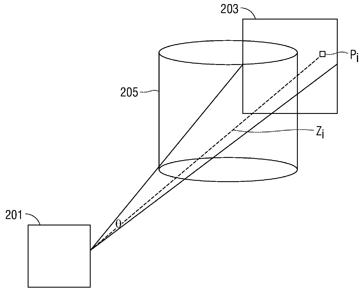
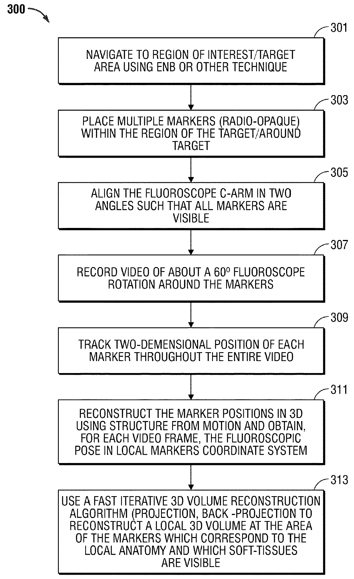
[0045]The present disclosure is directed to a system and method for constructing local three dimensional volumetric data, in which small soft-tissue objects are visible, from a video stream captured by a standard fluoroscopic imaging device available in most procedure rooms. The present disclosure is further directed to a system and method for determining a location of a medical device relative to a soft tissue target within a patient according to the three dimensional volumetric data. The constructed fluoroscopic-based local three dimensional volumetric data or the location of the medical device relative to the soft tissue target may be used for guidance, navigation planning, improved navigation accuracy, navigation confirmation, and treatment confirmation.

[0046]The terms “tool”, “surgery instrument”, “surgical device”, “energy device”, “medical device” and alike may be used hereby interchangeably.

[0047]FIG. 1 depicts an Electromagnetic Navigation (EMN) system 100 configured for re...


PUM


Abstract

A system for constructing fluoroscopic-based three-dimensional volumetric data of a target area within a patient from two-dimensional fluoroscopic images including a structure of markers, a fluoroscopic imaging device configured to acquire a sequence of images of the target area and of the structure of markers, and a computing device. The computing device is configured to estimate a pose of the fluoroscopic imaging device for at least a plurality of images of the sequence of images based on detection of a possible and most probable projection of the structure of markers as a whole on each image of the plurality of images. The computing device is further configured to construct fluoroscopic-based three-dimensional volumetric data of the target area based on the estimated poses of the fluoroscopic imaging device.

Description

CROSS-REFERENCE TO RELATED APPLICATIONS[0001]This application is a continuation-in-part of U.S. application Ser. No. 15 / 224,812, filed on Aug. 1, 2016 which claims priority to U.S. Provisional Application Ser. No. 62 / 201,750, filed on Aug. 6, 2015. The disclosure of each of the prior applications is considered part of and is incorporated by reference in the disclosure of this application.BACKGROUNDTechnical Field[0002]The present disclosure relates to a system, apparatus, and method of navigation, position confirmation and position correction for surgical procedures. More particularly, the present disclosure relates to a system and method for constructing a fluoroscopic-based three-dimensional volume from two dimensional fluoroscopic images captured using a standard fluoroscopic imaging device and for utilizing this fluoroscopic-based three dimensional volume for real-time determination of the location of a medical device relative to a target within a patient.Description of Related ...

Claims


Application Information

Patent Timeline
IPC IPC(8): A61B6/12A61B6/00A61B34/20
CPCA61B6/12A61B6/463A61B6/466A61B6/487A61B34/20A61B6/5235A61B2034/2065A61B2090/367A61B2090/3762A61B2090/3966A61B2090/3983A61B2034/2051A61B2090/3908A61B2034/2046A61B6/582A61B6/4441A61B6/0492A61B6/022A61B6/00
Inventor WEINGARTEN, OREN P.BARAK, RONBIRENBAUM, ARIELALEXANDRONI, GUY
Owner TYCO HEALTHCARE GRP LP
Why Eureka
Try Eureka