Confocal microscope with positionable imaging head

Active Publication Date: 2018-05-17
CALIBER IMAGING & DIAGNOSTICS
View PDF9 Cites 14 Cited by
  • Summary
  • Abstract
  • Description
  • Claims
  • Application Information

AI Technical Summary

Benefits of technology

[0004]Accordingly, it is an object of the present invention to provide a confocal microscope having an imaging head supported from a platform where the imaging head is positionable over the platform or beside the platform as desired for imaging tissue samples, thereby enabling ex-vivo imaging of tissue samples as may be disposed on a stage upon the platform and in-vivo imaging of tissue samples as may be disposed beside the platform.

Problems solved by technology

While useful for imaging in-vivo tissue, such as a skin lesion without removal from a patient, it is cumbersome when one wishes to image ex-vivo tissue samples as may be mounted on a microscope stage.

Method used


Image

  • Confocal microscope with positionable imaging head
  • Confocal microscope with positionable imaging head
  • Confocal microscope with positionable imaging head

Examples


Embodiment Construction

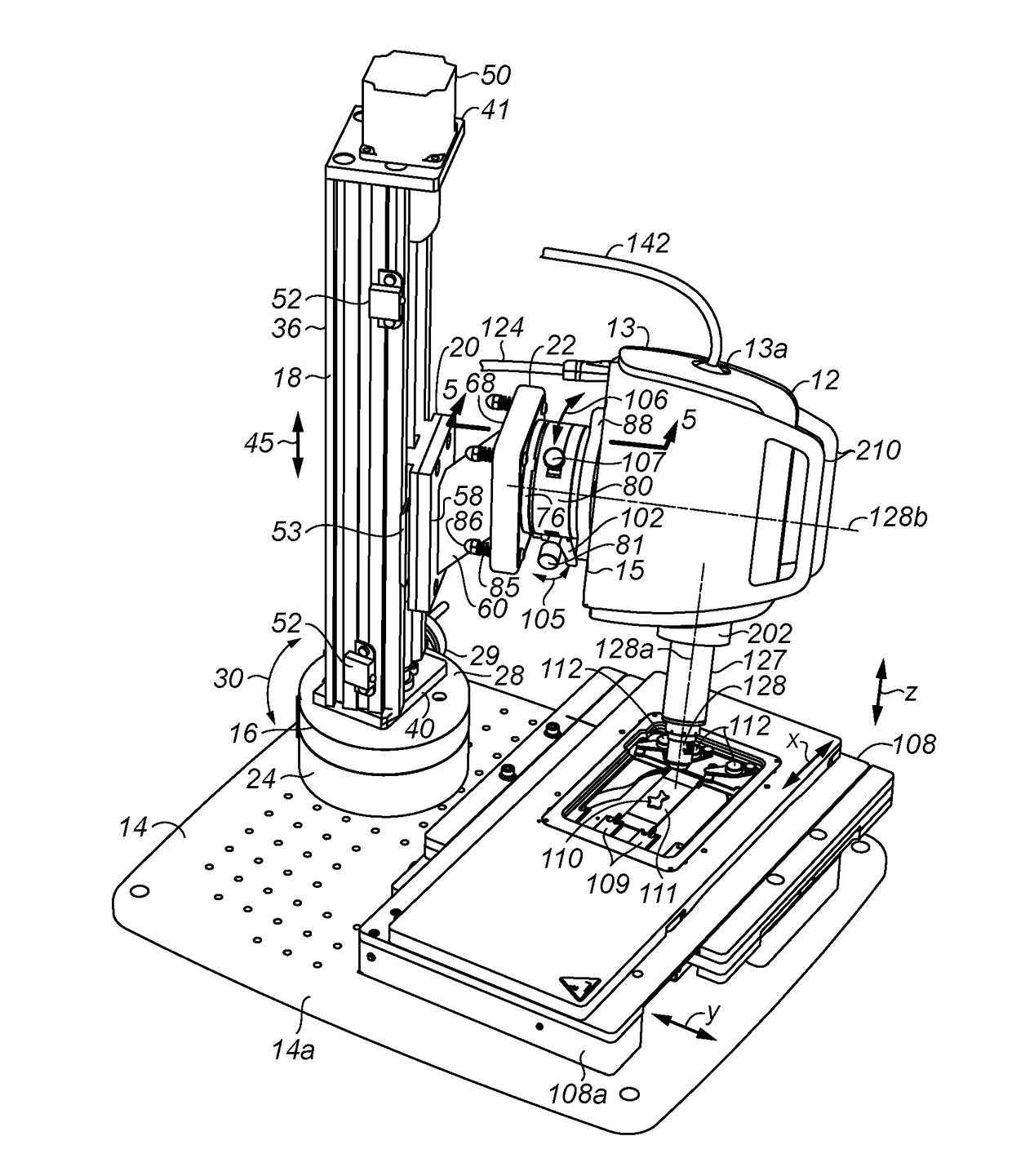
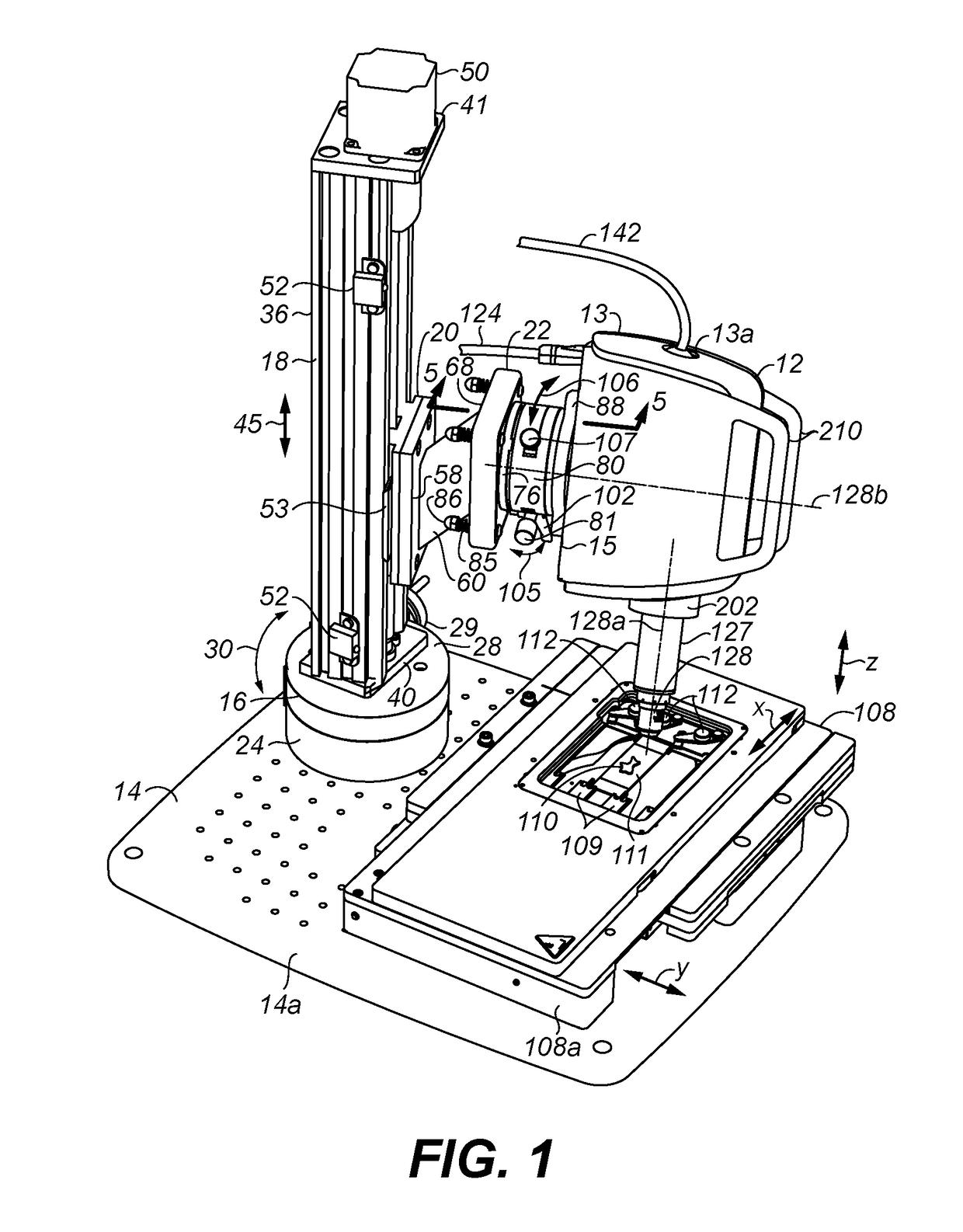
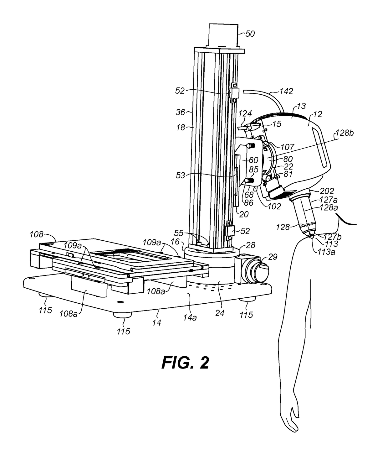
[0025]Referring to FIGS. 1, 2, and 3, an imaging head 12 of a microscope 10 (FIG. 8) is shown in a housing 13 supported over a platform (or base) 14 having an upper surface 14a along a horizontal or dimension or plane. A second (or rotary) stage 16 is mounted to platform 14 for rotating a first (or vertical) stage 18 about a vertical dimension, i.e., perpendicular to the horizontal dimension along which upper surface 14a of platform 14 extends. Vertical stage 18 is a vertically disposed linear slide stage which carries a movable carriage 20 for translation along such vertical dimension. Carriage 20 is coupled by a mounting arm 22 to a base 15 of housing 13 of imaging head 12. The mounting arm 22 enables adjustment of tilt and rotation of the imaging head 12 at a desired adjustable rotational position and height position as set by rotary stage 16 and vertical stage 18, respectively, as will be described later below in more detail.

[0026]The imaging head 12 has an optical system 11 for...


PUM


Abstract

A confocal microscope for imaging tissue having an imaging head for capturing optically formed microscopic sectional images of tissue samples, a platform upon which is disposed a linear stage for moving the imaging head along a vertical dimension, and a rotary stage to rotate the linear stage and imaging head about the vertical dimension. A mounting arm couples the imaging head to the linear stage to adjust tilt of the imaging head and to rotate the imaging head about a normal axis perpendicular to an optical axis of an objective lens of the imaging head. In a first mode of operation, the imaging head is positioned to image an ex-vivo or in-vivo tissue sample upon the platform, such as ex-vivo tissue sample mounted upon a movable specimen stage, and in a second mode of operation the imaging head is positioned to image an in-vivo tissue sample beside the platform.

Description

[0001]This application claims priority to U.S. Provisional Patent Application No. 62 / 421,270, filed Nov. 12, 2016, which is incorporated herein by reference.FIELD OF THE INVENTION[0002]The present invention relates to a confocal microscope having a positionable imaging head mounted to a platform, and particularly to, a confocal microscope having an imaging head mounted to a platform positionable in one mode to image tissue samples disposed upon the platform, such as an ex-vivo tissue specimen on a movable specimen stage, and in another mode to image tissue samples beside the platform, such as in-vivo skin tissue of large animals or humans.BACKGROUND OF THE INVENTION[0003]Confocal microscopes optically section tissue to produce microscopic images of tissue sections without requiring histological preparation of the tissue on slides (i.e., slicing, slide mounting, and staining). Such sectional images produced may be on or under the surface of the tissue. An example of a confocal micros...

Claims


Application Information

Patent Timeline
Patent Type & Authority Applications(United States)
IPC IPC(8): G02B21/26G02B21/24G02B21/00G02B21/36A61B5/00A61B90/20
CPCG02B21/26G02B21/24G02B21/248G02B21/0028G02B21/361G02B21/368A61B5/0059A61B90/20A61B2562/0233G02B21/0032G02B21/0076A61B5/0068A61B5/441A61B5/7225
Inventor CRAMB, ALLISON L.DISTASIO, CHRISTOPHER C.FOX, WILLIAM J.RIDGE, STEVENWATKINS, SIMON C.
Owner CALIBER IMAGING & DIAGNOSTICS
Why Eureka
Try Eureka