Methods for cell labeling and medical imaging
a cell labeling and medical imaging technology, applied in the field of ex vivo labeling of biological materials for in vivo imaging, can solve the problems of limiting the utility of cell tracking to shorter observational periods, poor in vivo imaging characteristics of radiotracers, and appreciable efflux of sequestered radioactivity after labeling
- Summary
- Abstract
- Description
- Claims
- Application Information
AI Technical Summary
Benefits of technology
Problems solved by technology
Method used
Image
Examples
example 1
Overview of Example 1
[0066]Background: With the recent growth of interest in cell-based therapies and radiolabeled cell products, there is a need to develop more robust cell labeling and imaging methods for in vivo tracking of living cells. This study in Example 1 describes evaluation of a novel cell labeling approach with the PET isotope 89Zr (T1 / 2=78.4 hours). Zr-89 may allow PET imaging measurements for several weeks and take advantage of the high sensitivity of PET imaging.
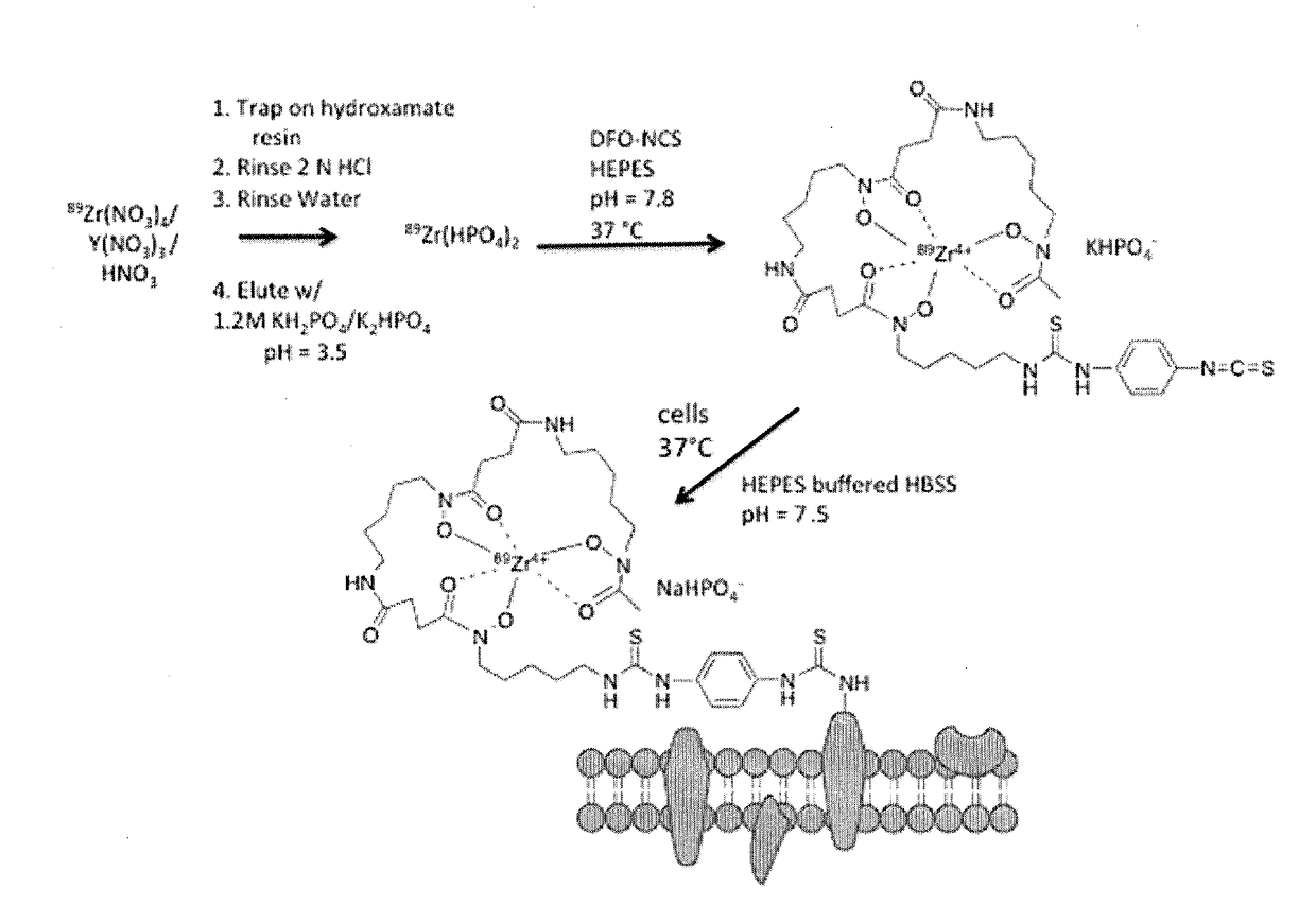
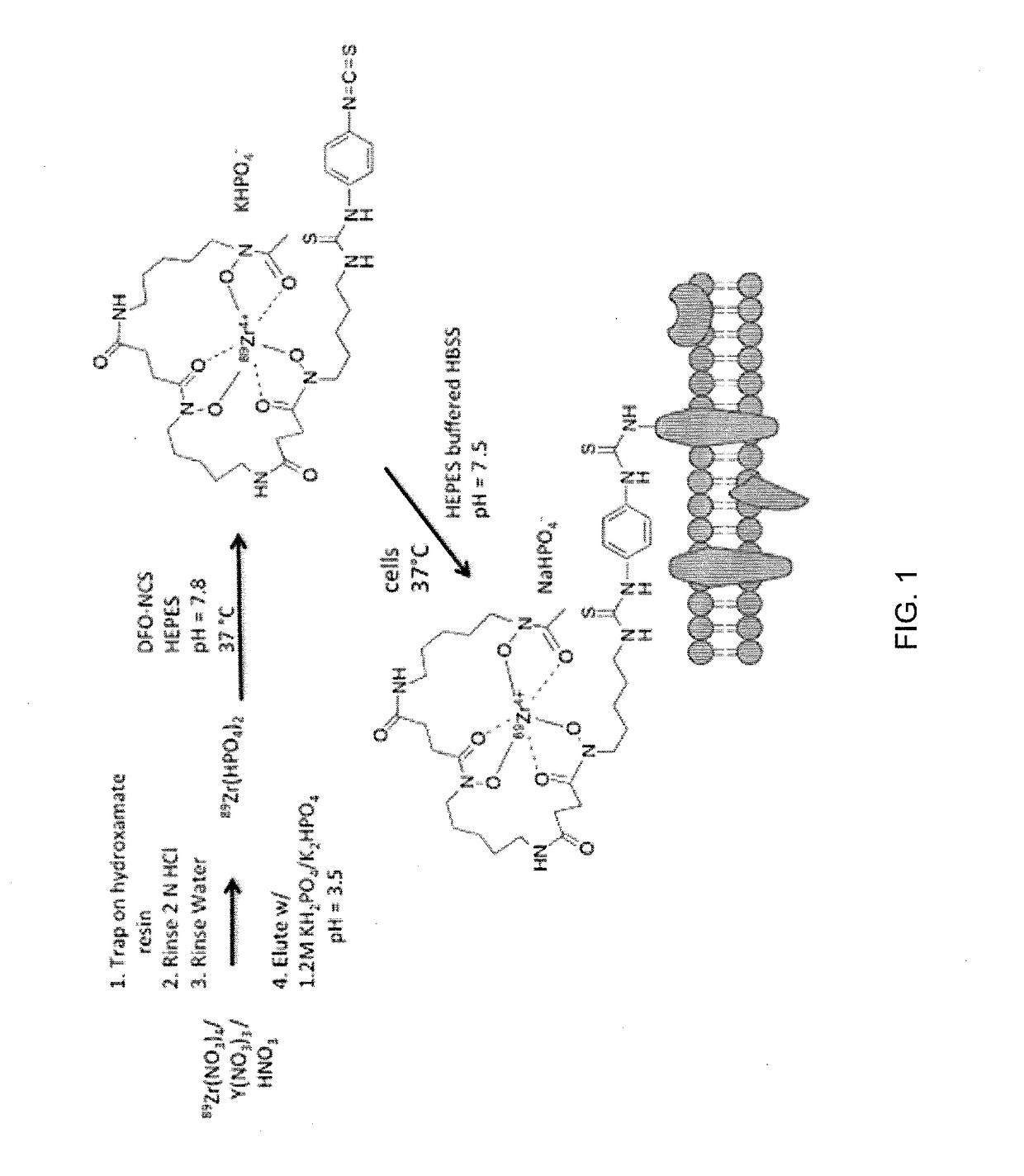
[0067]Methods: A novel cell-labeling agent, 89Zr-desferrioxamine-NCS (89Zr-DBN), was synthesized. Mouse derived melanoma cells (mMCs), dendritic cells (mDCs) and human mesenchymal stem cells (hMSCs) were covalently labeled with 89Zr-DBN via the reaction between the NCS group on 89Zr-DBN and primary amine groups present on cell surface membrane protein. The stability of the label on the cell was tested by cell efflux studies for 7 days. The effect of labeling on cellular viability was tested by proliferation, t...
example 2
[0097]In this Example, we report the synthesis and evaluation a novel virus labeling agent, 89Zr-desferrioxamine-NCS (89Zr-DBN) for non-invasive PET based in vivo tracking of administered virus.
Background
[0098]There is a growing interest in using viral based gene therapy for treating wide range of diseases. Viral based gene delivery agents for gene therapy hold tremendous promise but like drugs, a better understanding of their pharmacokinetics is critical to their advancement. This Example 2 study describes a novel viral labeling approach with 89Zr (T1 / 2=78.4 h) for potential PET imaging of viral biodistribution in the body.
Methods
[0099]A new viral labeling agent, 89Zr-desferrioxamine-NCS (89Zr-DBN), was synthesized. The adeno-associated viruses (AAVs) were covalently labeled with 89Zr-DBN via the reaction between the NCS group on 89Zr-DBN and primary amine groups present on capsid / envelope protein. The viral radiolabeling yield was quantified, following which cell binding and cell-...
example 3
[0115]In this Example, we report the tracking of transplanted Zirconium-89 (89Zr)-labeled mesenchymal stem cells (MSCs) serially with positron emission tomography for 21 days.
[0116]Human MSC Preparation: Human MSCs from healthy donors were obtained from the Human Cellular Therapy Laboratory, Mayo Clinic, Rochester, Minn., USA. These cells have been characterized with respect to surface markers. Briefly, they are CD73(+), CD90(+), CD105(+), CD44(+), and HLA-ABC(+), and they are being used in several clinical trials.
[0117]Green fluorescent protein (GFP) Transfection: MSCs were transfected with GFP lentivirus from Mayo Clinic Rochester Labs, Rochester, Minn., USA. MSCs were grown overnight in media containing the GFP lentivirus. The medium was changed to complete growth medium the next day, and cells were checked for fluorescence after 48 hours. Once fluorescence was confirmed, the cells were cultured in complete media that contained 1 μg puromycin per milliliter. Cells containing the ...
PUM
 Abstract
Description
Claims
Application Information
 - R&D
- Intellectual Property
- Life Sciences
- Materials
- Tech Scout
- Unparalleled Data Quality
- Higher Quality Content
- 60% Fewer Hallucinations
Browse by: Latest US Patents, China's latest patents, Technical Efficacy Thesaurus, Application Domain, Technology Topic, Popular Technical Reports.
© 2025 PatSnap. All rights reserved.Legal|Privacy policy|Modern Slavery Act Transparency Statement|Sitemap|About US| Contact US: help@patsnap.com


