Spinal subpial gene delivery system
a gene delivery and spinal cord technology, applied in the field of spinal cord subpial gene delivery system, can solve the problems of limited in vivo use of aav-based vectors to achieve gene-specific silencing or upregulation in the central nervous system
- Summary
- Abstract
- Description
- Claims
- Application Information
AI Technical Summary
Benefits of technology
Problems solved by technology
Method used
Image

Examples
example 1
Materials and Methods
[0085]Animals and General Surgical Preparation—
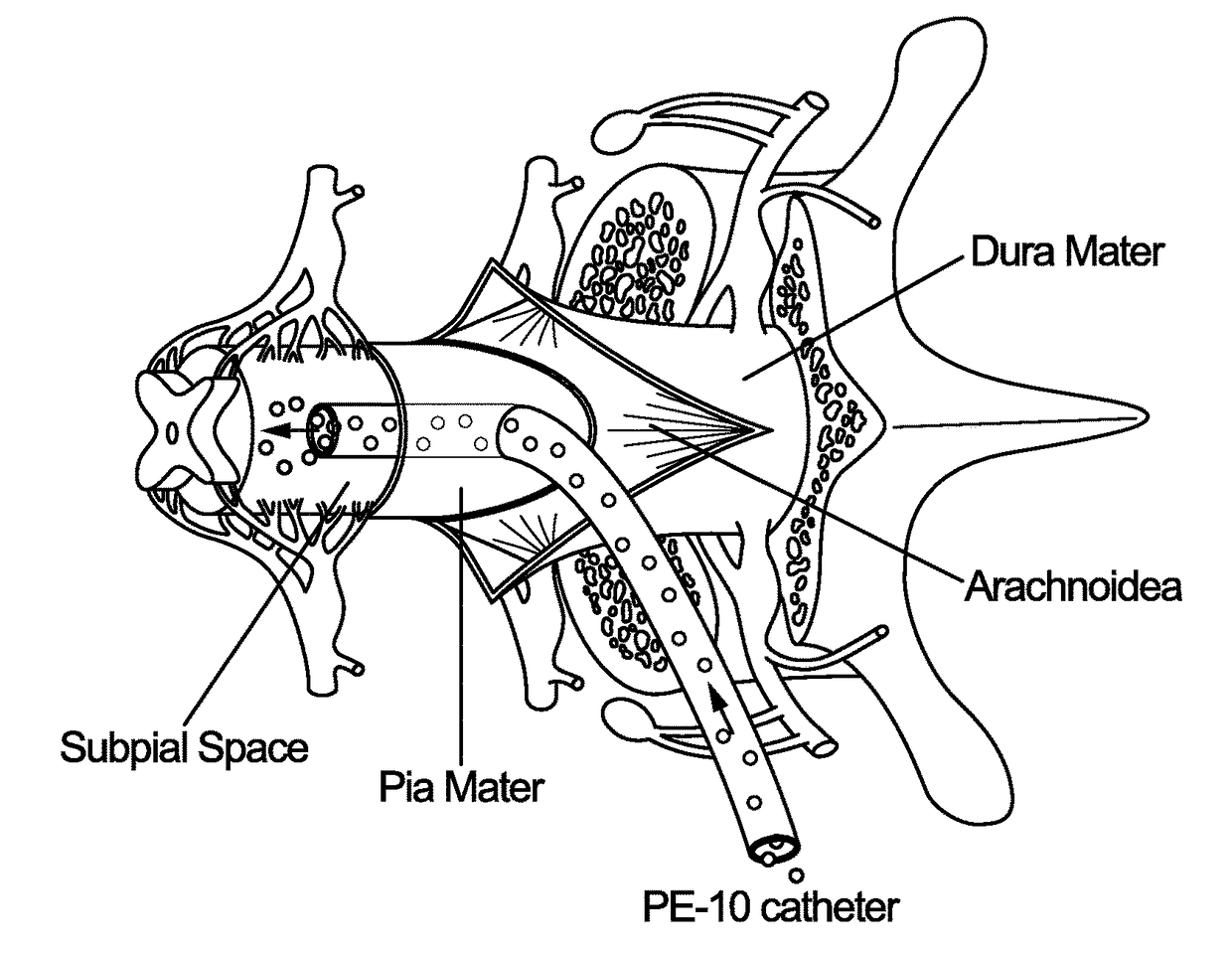
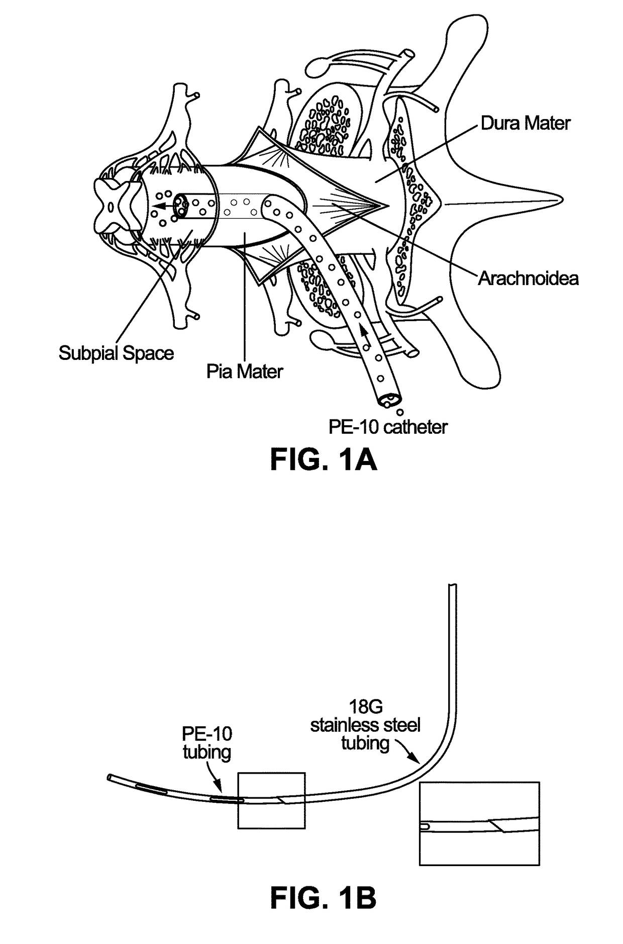
[0086]Adult Sprague-Dawley rats (male and female, 250-350 grams; n=16) or adult minipigs resulting from cross-breeding of Minnesota and Gottingen strains (both sexes; 30-40 kg; n=6) were used. Rats were anesthetized with 5% isoflurane and maintained at 2-3% of isoflurane during surgery depending on breathing rate and paw pinch response. The back of the rat was then shaved and cleaned with 2% chlorohexadine. After skin incision, the paravertebral muscle surrounding the cervical, thoracic or lumbar spinal vertebrae was removed and animals mounted into a spinal immobilization frame (Stoelting) using Cunningham's spinal clamps as previously described (Kakinohana, et al. (2004). Region-specific cell grafting into cervical and lumbar spinal cord in rat: a qualitative and quantitative stereological study. Experimental neurology 190: 122-132). To expose the spinal cord a dorsal laminectomy of corresponding vertebra was perf...
example 2
Parenchymal AAV9-Mediated Transgene Expression after Single Supial Bolus
[0096]Initially, the potency of single bolus subpial AAV9-UBI-GFP or AAV9-UBI-RFP delivery in rats and pigs was tested. Animals received 20 μl (rat) or 200 μl (pig) of AAV9 vector in 2.5% dextran solution delivered into subpial space of cervical (C4-6), thoracic (Th6-9) or lumbar (L2-L5) segments. At 6-8 weeks after AAV9 delivery spinal cords were dissected from 4% paraformaldehyde-fixed animals and imaged in situ using Avis fluorescence system. Transverse or horizontal spinal cord sections were then cut from AAV9-injected segments and analyzed for the presence of GFP or RFP and co-stained with neuronal (NeuN) and glial (GFAP) antibodies. In both rat and pig spinal cord an intense GFP or RFP expression was seen on the surface of the spinal cord and was readily identified by visual inspection as yellow-green or red areas. FIGS. 1H and 1J show the presence of RFP (red color) in transversely cut pig spinal cord and...
example 3
GFP Expression in Distant Spinal Segments
[0100]Next, the extent of descending spinal tract GFP expression in lumbar spinal cord was characterized after subpial injection of AAV9-UBI-GFP into the subpial space of mid-thoracic (Th6-7) or lower cervical segments in both rats and pigs. At 3-6 weeks after subpial AAV9 delivery, an intense GFP expression was seen throughout the whole lumbar spinal cord. Using transverse lumbar (L2-L6) spinal cord sections taken from pigs high intensity GFP expression in transversely-cut axons in the lateral and ventral funiculi was readily identified without additional GFP immunostaining (FIG. 4A, white asterisks). In these regions a similar density of GFP+ axons was seen throughout the whole white matter. In comparison to lateral and ventral funiculi, a relatively lower number of GFP+ axons was seen in the dorsal funiculi. (FIG. 4A, DF). Consistent with the degree of axonal labeling seen in the white matter of lateral and ventral funiculi a dense network...
PUM
| Property | Measurement | Unit |
|---|---|---|
| Fraction | aaaaa | aaaaa |
| Magnetic field | aaaaa | aaaaa |
| Disorder | aaaaa | aaaaa |
Abstract
Description
Claims
Application Information
 - R&D
- Intellectual Property
- Life Sciences
- Materials
- Tech Scout
- Unparalleled Data Quality
- Higher Quality Content
- 60% Fewer Hallucinations
Browse by: Latest US Patents, China's latest patents, Technical Efficacy Thesaurus, Application Domain, Technology Topic, Popular Technical Reports.
© 2025 PatSnap. All rights reserved.Legal|Privacy policy|Modern Slavery Act Transparency Statement|Sitemap|About US| Contact US: help@patsnap.com