
Methods for blood typing and antibody screening

a technology of applied in the field of blood typing and antibody screening, can solve the problems of false positive tests for hemagglutination, increase in the sedimentation rate of erythrocytes, and rouleaux formation, so as to prevent erythrocyte sedimentation, increase viscosity, and prevent erythrocyte sedimentation

Inactive Publication Date: 2017-11-02
T2 BIOSYST
View PDF4 Cites 2 Cited by
  • Summary
  • Abstract
  • Description
  • Claims
  • Application Information

AI Technical Summary

Benefits of technology

This patent describes different methods for preventing the red blood cells from sedimenting in blood samples. One method involves using fibrin mesh or a synthetic gel to suspend the red blood cells. Another method involves adding an additive to increase viscosity and prevent sedimentation. These methods may help to improve the quality and accuracy of blood sample analysis.

Problems solved by technology

They are therefore most frequently screened, as severe complications can result from improper transfusions involving RBCs with these antigens.
These formations occur when the plasma protein concentration is high, and they cause an increase in the erythrocyte sedimentation rate.
Rouleaux formation is the most common cause of false positive tests for hemagglutination.
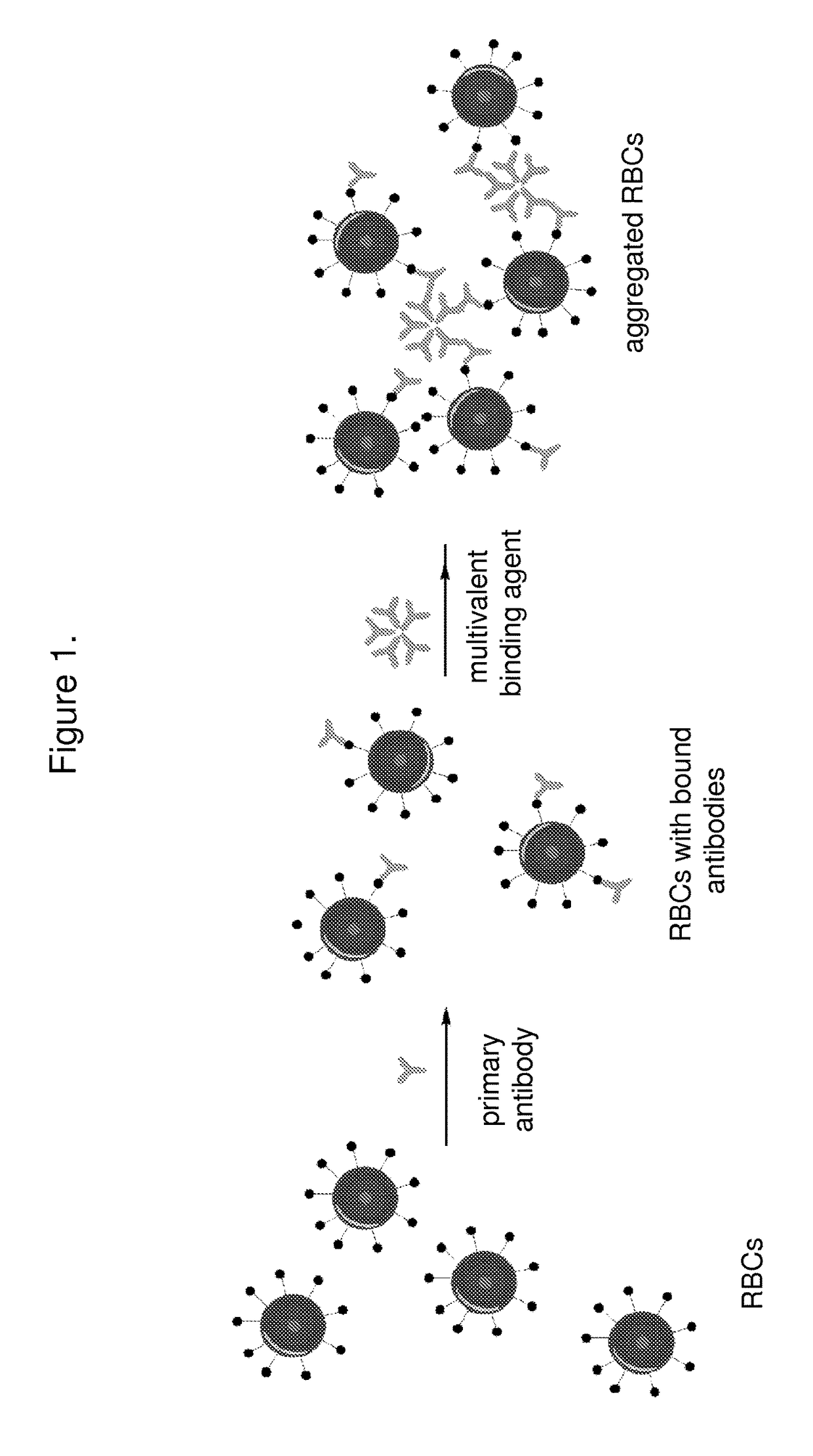
When IgM red blood cell antibodies are mixed with a suspension of erythrocytes bearing the corresponding antigens, each IgM molecule is large enough to simultaneously bind to red cell antigen sites on adjacent red cells resulting in hemagglutination.

Method used


Image

  • Methods for blood typing and antibody screening
  • Methods for blood typing and antibody screening
  • Methods for blood typing and antibody screening

Examples


example 1

Screening or Reverse Blood Typing

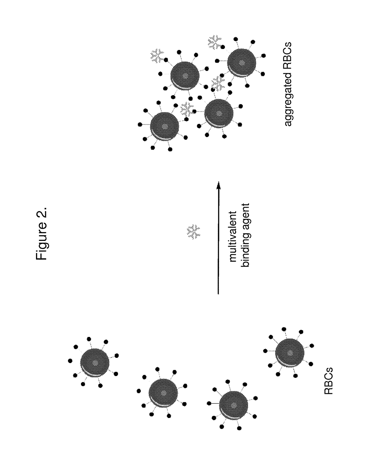
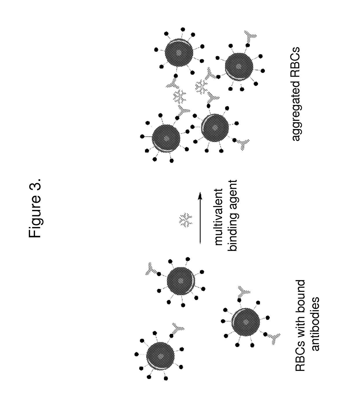
[0107]Briefly, the assay includes adding a small volume of reagent red blood cells (e.g. 10 μL) to a small volume (e.g. 10 μL) of patient serum or plasma. Potentiator can be added to accelerate the binding of antibodies to the surface of the reagent red blood cells. The sample can then be centrifuged and washed if desired. A multivalent binding agent such as monoclonal IgM can be added to promote aggregation. After mixing, the T2 reading can be taken to determine whether antibodies to the blood group on the reagent blood cells is present.

[0108]The principle of this assay is that T2MR is sensitive to RBC aggregation and can measure the rate of RBC aggregation. The extent of aggregation will depend on the binding of antibodies to the antigens on the surface of the RBCs and the subsequent complexation of the multivalent binding agent that cross-links antibodies on the surface of the RBCs. Aggregation is similar to clot formation, a process that can be w...

example 2

el Concept

[0110]This assay involves the comparison of two samples. One sample contains subject serum (e.g. 10 μL) and an equal volume of reagent RBCs and the other sample contains corresponding amounts of reagent RBCs combined with control serum lacking antibodies to the reagent RBCs. Comparison of the T2 signals of the target and control samples will occur over about 10 minutes. The change in T2 may either be the result of aggregation (presence of the settling peak, presence of a third peak, or change in the T2 of the settling peak relative to control).

example 3

ood Typing for ABOD Antigens

[0111]A small volume of washed patient red blood cells (e.g. 10 μL) are obtained and mixed separately with a small volume (e.g. 10 μL) of IgM against each of the ABOD antigens. After mixing, the T2 measurement is taken, possibly in a dynamic state with mixing. The T2 value can be compared to a standard curve or cut-off value to determine if the RBCs did indeed aggregate.


PUM


Abstract

The invention features systems and methods for the detection of analytes, particularly antigens, complement, and antibodies present in blood. The invention features a system for the detection of antigens, complement, and antibodies on the surface of RBCs and circulating antibodies in the blood.

Description

BACKGROUND OF THE INVENTION[0001]This invention features assays for the detection of analytes in blood and their use in medical treatment and diagnosis of disease.[0002]Blood is the circulating tissue of an organism that carries oxygen and nutritive materials to the tissues and removes carbon dioxide and various metabolic products for excretion. Whole blood consists of a pale yellow or gray yellow fluid, plasma, in which are suspended red blood cells (RBCs), white blood cells, and platelets. Red blood cells have proteins and sugars on their surfaces that have antigenic properties that can stimulate the production of antibodies.[0003]Blood testing for various antigens and antibodies is commonly performed in clinical laboratories. Specifically, the blood must be typed to determine the identity of antigens on the surface of the RBCs, and also determine whether there are any antibodies to erythrocyte antigens present in the serum or on the surface of RBCs. Traditionally blood typing, co...

Claims


Application Information

Patent Timeline
IPC IPC(8): G01N33/80G01N33/542
CPCG01N33/542G01N33/80G01N33/54373
Inventor LOWERY, JR., THOMAS JAYPAPKOV, VYACHESLAVMASSEFSKI, JR., WALTER W.
Owner T2 BIOSYST
Learn More