Method for automatic tissue segmentation of medical images

a tissue segmentation and medical image technology, applied in the field of images, can solve the problems of cumbersome and time-consuming, manual initialization of objects in 3d images, and inability to be reused

Active Publication Date: 2017-07-27
RIVERSIDE RES INST +1
View PDF0 Cites 15 Cited by
  • Summary
  • Abstract
  • Description
  • Claims
  • Application Information

AI Technical Summary

Benefits of technology

[0010]The proposed methods presented by Ishikawa, Delong and Boykov require seeds. Methods using a convex energy functional compared to methods using a non-convex energy functional (i.e., requiring initialization) can be more accurate and more flexible to achieve satisfactory results. Initializations (e.g., initial seeds) are not reusable, cumbersome and time consuming to initialize objects manually in a 3D image. In contrast, parameters used in a convex energy functional can be trained using typical images in each application setting, which are then applicable to all images in the same setting.
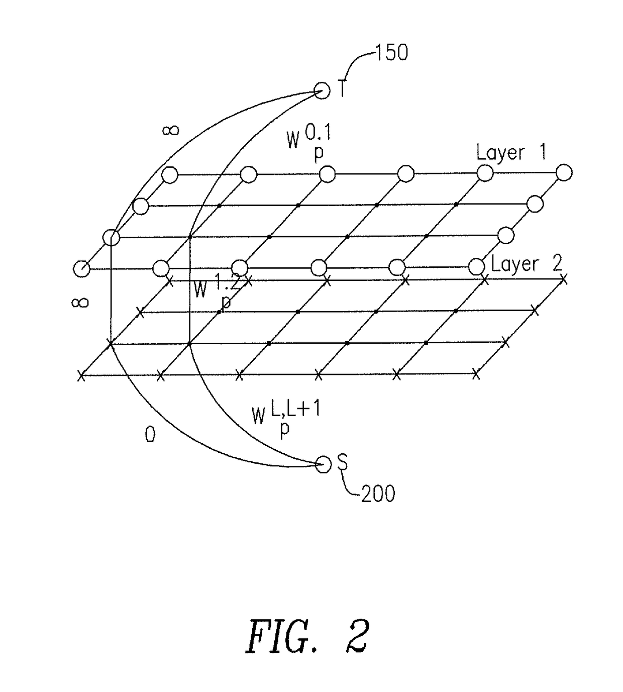
[0012]For segmenting nested objects, the present invention uses undirected links between adjacent layers. Therefore, the graph cut calculation is accelerated using fast max-flow algorithms for undirected graphs;
[0013]Second, it includes padded nodes with infinite-cost links to the sink, so that all cuts at the image boundary in each layer are costly. With these padded nodes, NGC can avoid incorrect segmentations even though they satisfy a containment relationship;
[0015]a multi-resolution approach is provided for solving the NGC to reduce the computation time.

Problems solved by technology

Shape preservation and distribution estimation are two critical problems for segmentation.
Initializations (e.g., initial seeds) are not reusable, cumbersome and time consuming to initialize objects manually in a 3D image.

Method used


Image

  • Method  for automatic tissue segmentation of medical images
  • Method  for automatic tissue segmentation of medical images
  • Method  for automatic tissue segmentation of medical images

Examples


example 1

[0049]3D Quantitative ultrasounds were used to detect cancerous regions in freshly dissected human lymph nodes (LNs). The LNs contain three parts—LN-parenchyma (LNP), fat and phosphate-buffered saline (PBS). The present invention was used to effectively detect the nested structure of the LN images.

[0050]Deep LNP pixels having a very dark intensity similar to PBS are recognized in the first iteration of NGC. The mean and variance of both the LNP cluster and the PBS cluster, resulting from K-means to determine Wp0.1(Ip) in (2), are used and the one having the smaller weight is chosen. This helps to make deep LNP pixels be segmented correctly.

[0051]FIG. 4(a) shows the original image obtained having the LNP, fat and PBS. FIG. 4(b) shows the segmentation of FIG. 4(a) after the first iteration of NGC using the initial distributions, where the deeper surrounding fat is falsely segmented as LNP because of attenuation effects. FIG. 5(a) shows the mean intensity of LNP (the line represented b...

example 2

[0054]The 3D HFU mouse-embryo images were acquired with a custom 40-MHz, five-element, annular-array system. For this study, 40 mouse-embryo 3D images spanning five mid-gestational stages from E10.5 to E14.5 were used. The dimension of the 3D image increases depending on gestational stages, ranging from by 149 by 40 to 200 by 200 by 100 voxels. The voxel size of the 3D HFU datasets was 25 by 25 by 50 / MI. For all 40 images, semi-automatic segmentations of brain ventricles (BVs) were available, while only 28 out of the 40 images had manual segmentations of the embryonic head available. The contour of the embryonic head was manually drawn using Amira visualization software by a small-animal ultrasound imaging expert. These manual segmentations were used as ground truth to quantify the performance of the present invention.

[0055]Accurate segmentation of the BV and the head is essential for assessing the normal and abnormal development of the central nervous system of mouse embryos. Table...


PUM


Abstract

A method to segment images that contain multiple objects in a nested structure including acquiring an image; defining the multiple objects by layers, each layer corresponding to one region, where a region contains an innermost object and all the objects nested within the innermost object; stacking the layers in an order of the nested structure of the multiple objects, the stack of layers having at least a top layer and a bottom layer; extending each layer with padded nodes; connecting the top layer to a sink and the bottom layer to a source, wherein each intermediate layer between the top layer and the bottom layer are connected only to the adjacent layer by undirected links; and measuring a boundary length for each layer.

Description

PRIORITY AND RELATED APPLICATION[0001]This application claims priority to U.S. Provisional Patent Application No. 62 / 281,271 filed Jan. 21, 2016 entitled “Method of Automatic Tissue Segmentation of Medical Images” and is hereby incorporated by reference in its entirety.FIELD OF THE INVENTION[0002]The present invention relates to the study of images derived from data acquired via Ultrasound, MRI, CT or Quantitative ultrasound, for example. More specifically the present invention relates to the segmentation of images (2D or 3D) that contain multiple objects in a nested structure.BACKGROUND OF THE INVENTION[0003]High-frequency (i.e., >15 MHz) ultrasound (HFU) provides fine-resolution images on the order of 100 um because of its short wavelengths (i.e., <100 um) and small focal-zone beam diameters. HFU is able to acquire 3D data in a manner of minutes with the potential to provide real time dynamic information. Because of its rapid data acquisition and commercial availability, HFU...

Claims


Application Information

Patent Timeline
Patent Type & Authority Applications(United States)
IPC IPC(8): G06T7/11G06T7/13G06T7/00
CPCG06T7/11G06T7/13G06T7/0012G06T7/162G06T2200/04G06T2207/10081G06T2207/10088G06T2207/10132G06T2207/30101
Inventor KUO, JEN-WEIMAMOU, JONATHANZHAO, XUANKETTERLING, JEFFREY A.ARISTIZABAL, ORLANDOTURNBULL, DANIEL H.WANG, YAO
Owner RIVERSIDE RES INST
Why Eureka
Try Eureka