
Method for Reduction of Artifacts and Preservation of Soft Tissue Contrast and Conspicuity of Iodinated Materials on Computed Tomography Images by means of Adaptive Fusion of Input Images Obtained from Dual-Energy CT Scans, and Use of differences in Voxel intensities between high and low-energy images in estimation of artifact magnitude

a computed tomography and soft tissue technology, applied in image enhancement, image analysis, instruments, etc., can solve the problems of deterioration of soft tissue contrast, marked deformation of artifacts near metallic or dense objects, and inability of conventional ct scanner detector elements to distinguish between x-ray photons of different energies, etc., to achieve fast approximation

Inactive Publication Date: 2017-04-20
RADIOLOGY UNIVERSE INST
View PDF9 Cites 2 Cited by
  • Summary
  • Abstract
  • Description
  • Claims
  • Application Information

AI Technical Summary

Benefits of technology

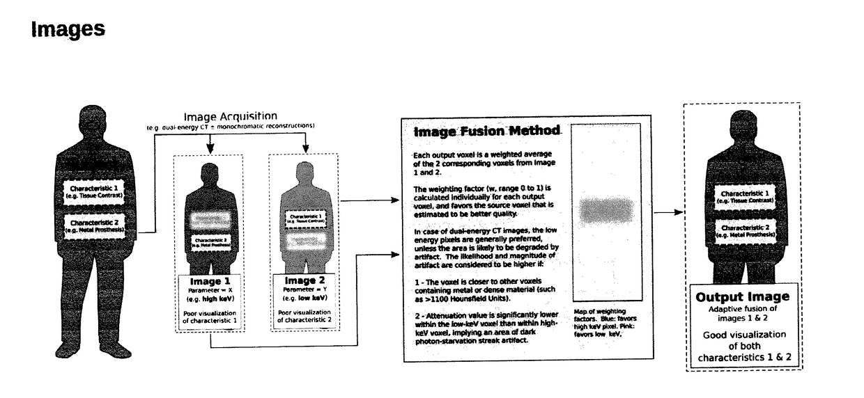
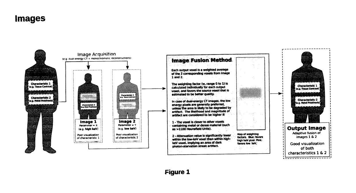
The present invention provides an improved method for reducing artifacts in computed tomography (CT) images, particularly those caused by metallic streak, beam hardening, and photon starvation. This is achieved by using voxel-by-voxel weighting factors that vary over the image region, rather than being constant for the entire input image. The new method also allows for maximum preservation of soft tissue contrast and conspicuity of iodinated materials on CT images. The proximity effect, which is akin to a gravitational field arising from dense materials, is estimated by calculating a quantity termed Metal Effect and is utilized to determine the weighting factors. To avoid sharp transitions between neighboring output pixels, a sigmoid curve or similar is used to limit the value of weighting factor. Overall, this method produces an image that is closer to the natural appearance of the patient's body, allowing for better visualization of iodinated contrast materials.

Problems solved by technology

Conventional CT scanner detector elements were typically unable to distinguish between x-ray photons of differing energies.
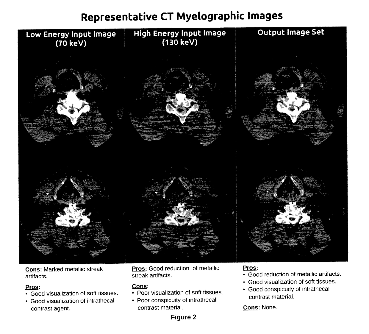
Regardless of acquisition method, typically the low-energy CT images (whether poly- or mono-monochromatic) exhibit relatively good visualization of soft tissues and iodinated contrast, but are markedly degraded by artifacts near metallic or dense objects.
Although raising photon energy leads to reduction metallic and beam hardening artifacts, such methods inversely result in degradation of soft tissue contrast and reduction in conspicuity of iodinated contrast agents.
Using such intermediate image may produce a reasonable compromise trade-off between artifacts vs. soft tissue visualization, but such a resultant intermediate image neither excels in artifact suppression nor soft tissue contrast and iodinated contrast material conspicuity.

Method used


Image

  • Method for Reduction of Artifacts and Preservation of Soft Tissue Contrast and Conspicuity of Iodinated Materials on Computed Tomography Images by means of Adaptive Fusion of Input Images Obtained from Dual-Energy CT Scans, and Use of differences in Voxel intensities between high and low-energy images in estimation of artifact magnitude
  • Method for Reduction of Artifacts and Preservation of Soft Tissue Contrast and Conspicuity of Iodinated Materials on Computed Tomography Images by means of Adaptive Fusion of Input Images Obtained from Dual-Energy CT Scans, and Use of differences in Voxel intensities between high and low-energy images in estimation of artifact magnitude
  • Method for Reduction of Artifacts and Preservation of Soft Tissue Contrast and Conspicuity of Iodinated Materials on Computed Tomography Images by means of Adaptive Fusion of Input Images Obtained from Dual-Energy CT Scans, and Use of differences in Voxel intensities between high and low-energy images in estimation of artifact magnitude

Examples


Embodiment Construction

[0020]Included C++ code excerpts from an example embodiment are intended to convey the underlying methods to those skilled in the art. Although the reference code processes dual-energy CT images, essentially the same method, or some of the provided computer code, can be used to process images obtained via other modalities, such as MRI, to optimize other characteristics, such as reduction of motion artifact (as opposed to streak artifact), or increase conspicuity of pathologic processes or normal structures. Third image (or more) can be optionally incorporated into this method by including additional terms in the voxel-by-voxel weighted average described above. Such a third image can also be used as a determinant of weight (w) itself (similar or substituted for the aforementioned Metal Effect image, which is calculated rather than acquired in the example embodiment). A voxel-by-voxel parameter similar to (w) above, can also be used to represent a target monochromatic energy level rat...


PUM


Abstract

Image processing method is presented along with reference source code and sample outputs. This method processes 2 (or more) input images which were obtained at different photon energies (or differing in another parameter), and produces an output image that emphasizes the best characteristics of each of the input images, while suppressing the undesired characteristics of each input image. This is different from prior dual-energy CT methods which produce an intermediate compromise image, with an intermediate soft tissue contrast and intermediate artifact suppression, and also different from prior methods that sacrifice soft tissue contrast to maximize artifact reduction.

Description

Field of the Invention[0001]The invention relates to the fields of image processing, radiology images, in particular an improved image reconstruction method from dual-energy Computed Tomography (CT) images. Same technique can be also be applied to other modalities such as MRI, or photographic images.Description of Prior Art[0002]Computerized Tomography (CT) has been extensively used in the fields of radiology, medical diagnosis, industrial quality assurance, and security systems. Conventional CT scanners typically emit a polyenergetic beam consisting of a wide spectrum of differing x-ray photon energies, with the energy spectrum adjustable by the operator of the scanner, typically by setting the acceleration voltage or changing beam filtering materials. Such adjustments may be desired to optimize the resultant image based on considerations such as girth of the imaged structure, presence of metallic implants or other dense material, use of iodinated contrast materials, and patient ra...

Claims


Application Information

Patent Timeline
IPC IPC(8): G06T11/00G06T7/00
CPCG06T11/008G06T7/0002G06T2207/10088G06T2207/10081G06T2207/30168
Inventor KULON, MICHAL
Owner RADIOLOGY UNIVERSE INST
Learn More