
Magnetic apparatus and methods of use

a magnetic device and magnetic technology, applied in the field of magnetic devices and methods, can solve the problems of critical lack of disease-modifying oa drugs (dmoad), biochemical changes detectable, and chronic inflammation that contributes to joint destruction

Active Publication Date: 2016-12-01
UNIV OF FLORIDA RES FOUNDATION INC
View PDF5 Cites 0 Cited by
  • Summary
  • Abstract
  • Description
  • Claims
  • Application Information

AI Technical Summary

Benefits of technology

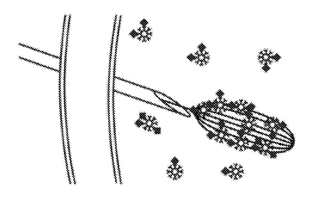
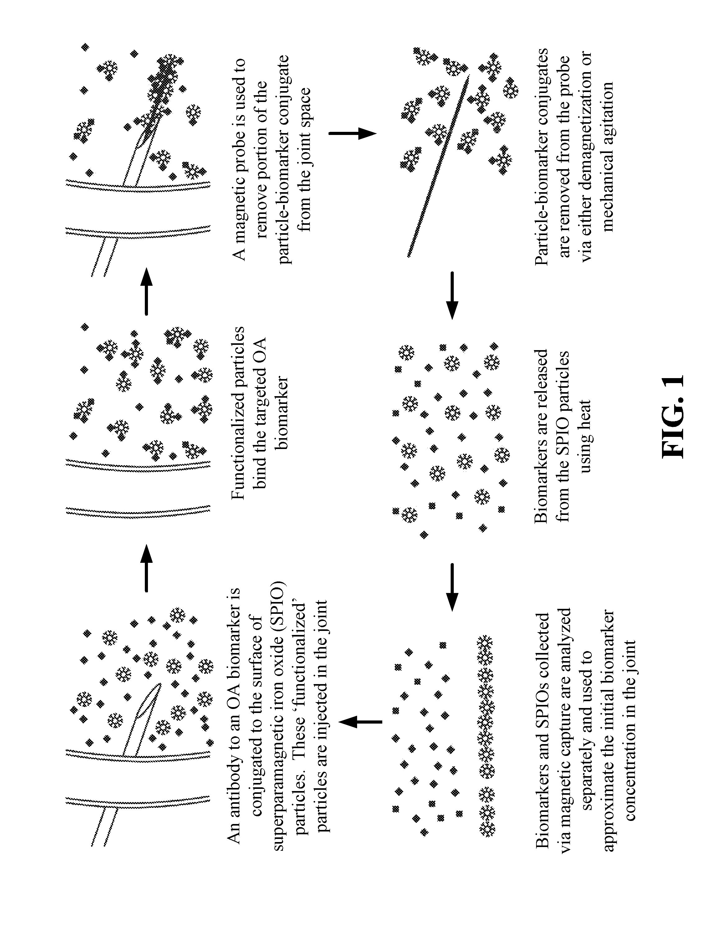
The patent text describes new methods and compositions for treating inflammation in joints. These methods involve using magnetic micro / nanoparticles to capture inflammatory mediators from the inflamed area. The particles are injected into the joint and a magnetic needle is used to collect them. The collected particles can then be analyzed to measure the severity of the inflammatory response. The method is safe and effective, as it allows for the removal of key inflammatory mediators without the need for removing synovial fluid. The patent also describes the use of targeted particle functionalization and the extraction of previously injected particles from a joint for analysis. Overall, the patent provides new tools for the diagnosis and treatment of joint inflammation.

Problems solved by technology

These mediators are part of the wound healing response, but if up-regulated chronically, these mediators can cause a chronic inflammation that contributes to joint destruction.
There is a critical lack of disease modifying OA drugs (DMOAD), due in part to an inability to clinically detect early stage OA.
Though promising biomarkers in urine and serum have been identified through the OA Biomarkers Global Initiative, biochemical changes detectable in the urine or serum are not specific to an affected joint, are dilute relative to levels within the affected joint, and may not be detectable at the earliest stages of OA.
Harvesting biomarkers from the synovial fluid of the knee joint has proven to be challenging.
In particular, removal of synovial fluid is difficult because it is an incredibly viscous fluid that usually requires a large needle for its removal.
Use of a large needle is difficult in intermediate joints and impossible in smaller joints such as the metacarpalphalanges and facet joints.
Removal of synovial fluid from large joints such as the knee or hip, while easier, is still problematic due to a fairly high rate of “dry taps” or inability to remove fluid from those joints.

Method used


Image

  • Magnetic apparatus and methods of use
  • Magnetic apparatus and methods of use
  • Magnetic apparatus and methods of use

Examples


example 1

Manufacture of Magnetic Needle Tip

[0049]A magnetic needle tip was laser-machined from an NdFeB permanent magnet using a diode-pumped solid-state laser operating at a normal 10-15 picosecond pulse width and 355 nm wavelength. An 800-micron-thick disc of NdFeB was placed under the laser, and the laser cut down through the thickness of the disc starting with a 50×50 micron square at the top and reaching a base of approximately 500×500 microns. This resulted in a pyramidal shape with a height of approximately 800 microns and a tip width of approximately 50 microns.

[0050]During the above process, the laser remains nearly perpendicular to the magnet. The pyramidal shape is achieved by using the aspect ratio (shadowing effect). The laser starts at the top of the magnet with a very small square (shown in dark grey in the middle of the “X” in FIG. 4), and as the laser cuts deeper into the magnet, the square expands due to aspect ratio limitations. By the time the laser cuts all the way throu...

example 2

Determination of Efficiency of Magnetic Particle Harvesting

[0052]Microneedles were placed in a solution of polystyrene / iron oxide composite fluorescent superparamagnetic particles with approximately 30,000 particles / μL (1 μm particle diameter). Particles collecting on a magnetic needle can be seen in frame grabs from a video. In this experiment, microneedles were able to collect 100,000-900,000 microparticles from the solution. FIG. 5 shows the results of this assay, which demonstrate that NdFeB magnetic microneedles can collect polymeric particles with SPIONs embedded within the particle core.

example 3

Time Dependencies for Binding and Release of the CTX-II Biomarker to / from Anti-CTX-II Conjugated Magnetic Particles

[0053]Approximately 106 million of anti-CTX-II conjugated magnetic particles (approximately 220 antibody molecules per particle) were mixed with ether non-treated or hyaluronidase-treated bovine synovial fluid (SF) and subject to constant gentle mixing (final volume 900 μl). FIG. 6 shows that after 2.5 hours (6B) or indicated time intervals (6A), 100 μl aliquots were taken for analysis.

[0054]Particles were separated from synovial fluid by centrifugation. Pellets were washed with PBS containing 0.05% TWEEN-20, 2% BSA, and 2 mM EDTA for 1 minute (6A) or for indicated amounts of time (6B). Then bound CTX-II was released from the particles by heating for 3 minutes at 85° C. (see control experiment shown in FIG. 9). Samples were analyzed for CTX-II using Serum Pre-Clinical CartiLaps ELISA kit. These results show no dependence on the synovial fluid viscosity.


PUM

PropertyMeasurementUnit
heightaaaaaaaaaa
heightaaaaaaaaaa
widthaaaaaaaaaa

Abstract

Provided herein is a magnetic apparatus for collecting superparamagnetic particles from a subject. The superparamagnetic particles are previously injected into the subject and have ligands bound thereto that are specific for one or more non-cell biomarkers. In one embodiment, the superparamagnetic particles are injected into and retrieved from a cavity such as a joint cavity. These compositions and methods allow for the sequestration and removal of inflammatory mediators, as both a diagnostic of the local immune response and a therapeutic that can reduce inflammation in the local disease environment.

Description

CROSS-REFERENCE TO RELATED APPLICATION[0001]This application is a divisional and claims priority to U.S. Non-Provisional Patent Application entitled: “MAGNETIC APPARATUS AND METHODS OF USE, having Ser. No. 13 / 967,571, filed on Aug. 15, 2013, which claims priority to and the benefit of U.S. provisional application entitled “MAGNETIC APPARATUS AND METHODS OF USE”, having Ser. No. 61 / 863,267, filed Aug. 15, 2012, both of which are hereby incorporated by reference in their entirety.STATEMENT REGARDING FEDERALLY SPONSORED RESEARCH OR DEVELOPMENT[0002]This invention was made with Government support under Contract No.: R00AR057426 awarded by the National Institutes of Health. The Government has certain rights in this invention.BACKGROUND OF THE INVENTION[0003]In damaged tissues, inflammatory mediators (such as cytokines or proteolytic enzymes) are up-regulated. These mediators are part of the wound healing response, but if up-regulated chronically, these mediators can cause a chronic infla...

Claims


Application Information

Patent Timeline
Patent Type & Authority Applications(United States)
IPC IPC(8): A61B10/02G01N33/543G01N33/68A61B17/52A61B17/34
CPCA61B10/02A61B17/52A61B17/3421G01N2800/105G01N33/54326G01N2333/78G01N33/6887A61B10/00A61B10/0045G01N2800/102Y10T29/49
Inventor ALLEN, KYLE DOUGLASDOBSON, JON PAULYARMOLA, ELENA GEORGINAKAUFMAN, ZACHARYARNOLD, DAVID P.
Owner UNIV OF FLORIDA RES FOUNDATION INC
Learn More