Method and system for tomosynthesis projection images enhancement

Active Publication Date: 2016-06-30
GENERAL ELECTRIC CO
View PDF11 Cites 12 Cited by
  • Summary
  • Abstract
  • Description
  • Claims
  • Application Information

AI Technical Summary

Benefits of technology

The patent describes a method for improving the quality of tomosynthesis images by filtering them for different frequency bands to account for different sizes of objects such as calcifications, fibers, and glands, and to remove undesired artifacts. The method also includes denoising the tomosynthesis images prior to improving their quality. A successive display is provided that allows the user to quickly appreciate the breast content at different angulations, with an adjustable speed. Overall, the method improves the reliability and accuracy of tomosynthesis images.

Problems solved by technology

One problem to be addressed is that images acquired during tomosynthesis through digital receptors may be contaminated by a variety of noise sources.
One drawback is that a single tomosynthesis projection image at a given orientation or x-ray source position is very noisy because the dose per projection is not enough to be compared to a known 2D mammogram acquisition.

Method used


Image

  • Method and system for tomosynthesis projection images enhancement
  • Method and system for tomosynthesis projection images enhancement
  • Method and system for tomosynthesis projection images enhancement

Examples


Embodiment Construction

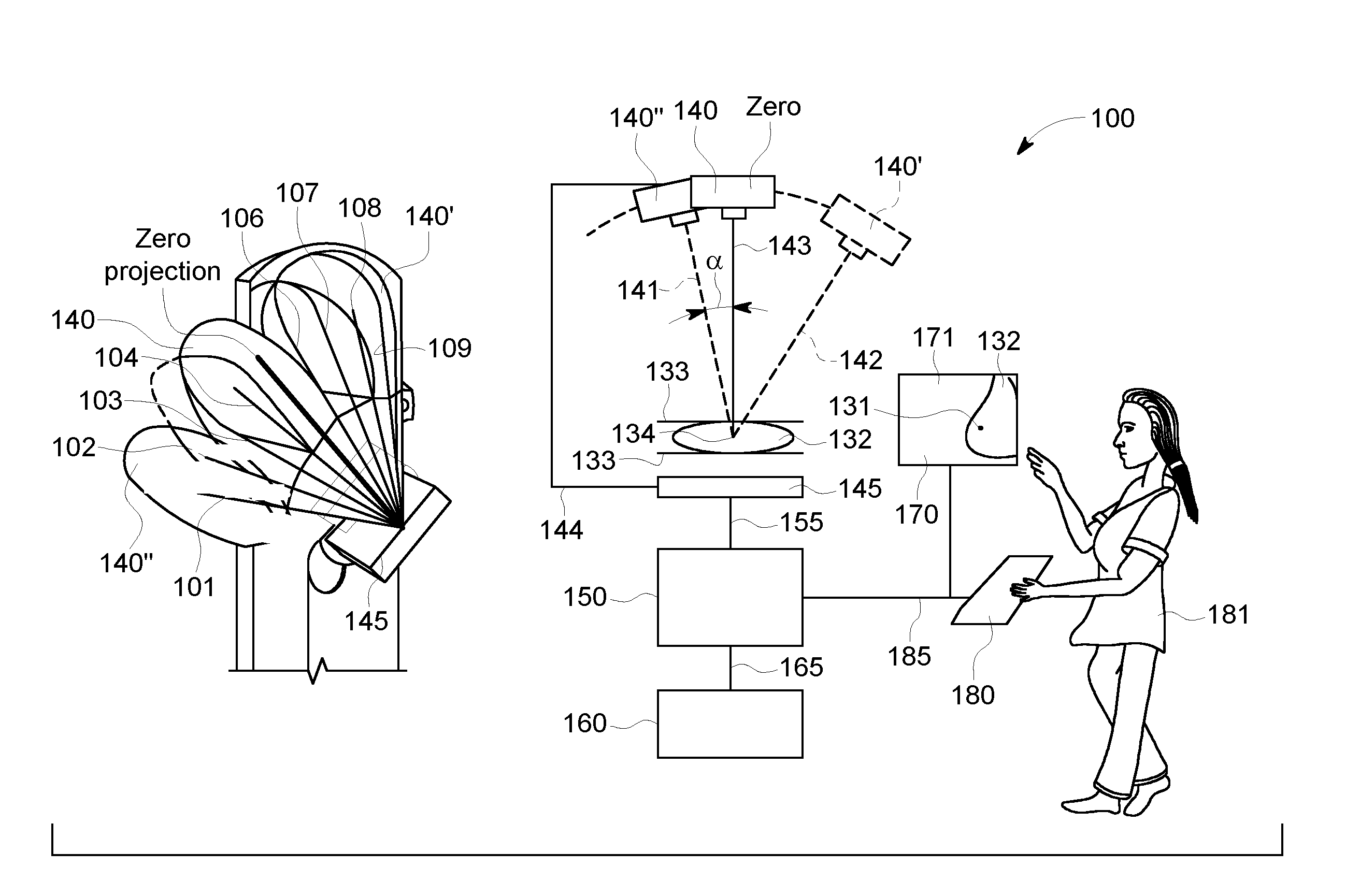
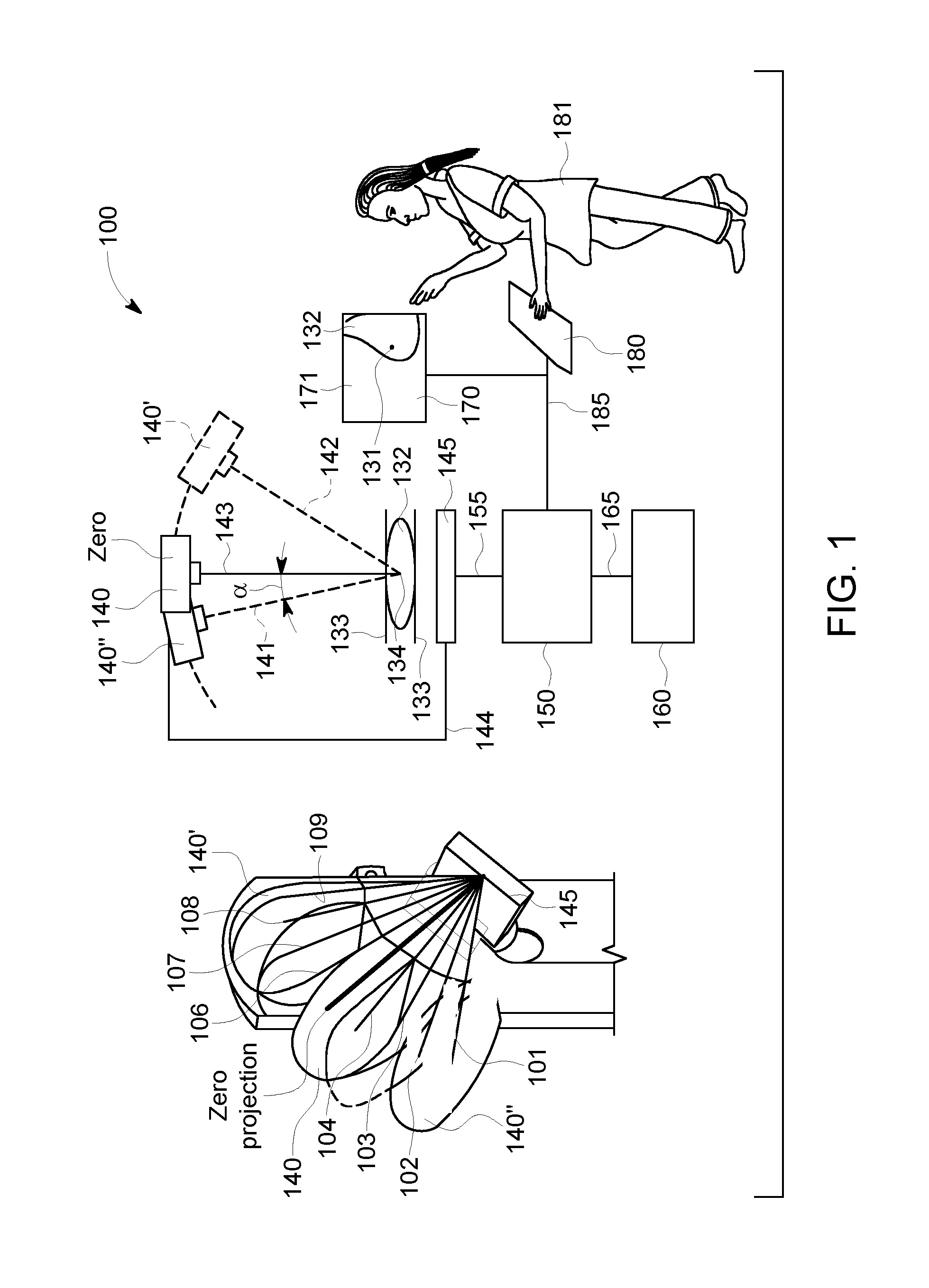
[0030]FIG. 1 is a diagrammatic illustration 1 of a system for obtaining an enhanced projection image of an object of interest, wherein the system 100 comprises a x-ray beam source 140 facing the detector 145. The x-ray beam source 140 and the detector 145 are connected by an arm 144. Between the detector 145 and the source 140 an object of interest 132 can be placed. In the system illustrated, the x-ray source 140 moves in an arc above a single detector 145. The detector 145 and a plurality of positions of the x-ray source 140′ and 140″ following an arc (see dashed line) are shown with dashed / solid lines and in a perspective partial view. In the shown arrangement, the detector 145 is fixed at the shown position and only the x-ray source 140 moves. The angle α is a projection angle enclosed by the zero-orientation and any other orientation such as 141 and 142. In this way multiple different views of the breast tissue can be acquired via the at least one x-ray source 140. The projecti...


PUM


Abstract

Method and system for obtaining images of an object of interest using a system comprising an x-ray source facing a detector. The method and system enable the acquiring of a plurality of 2D projection images of the object of interest in a plurality of orientations. A selected 2D projection image such as the zero projection of the plurality of projections can be enhanced by using at least a subset of the plurality of tomosynthesis projection images. The obtained enhanced 2D projection image is displayed for review.

Description

BACKGROUND[0001]The present disclosure relates to the field of tomosynthesis and to a method and system for processing tomosynthesis imaging data for obtaining enhanced projection images.[0002]X-ray imaging systems have become a valuable tool in medical applications such as for the diagnosis of many diseases. As standard screening for breast cancer mammography 2-dimensional (2D) x-ray images are taken across the entire breast tissue. These known 2D mammograms are limited by tissue superimposition. That is to say, lesions may be masked by the tissue above or underneath, or normal structures may mimic a lesion. In order to minimize limitations of standard 2D-mammography caused by tissue superimposition, digital breast tomosynthesis using digital receptors has been developed.[0003]The tomosynthesis systems employ at least one x-ray tube, which is moved in an arc above a stationary detector. In digital breast tomosynthesis (DBT) the volume information of an object of interest can be der...

Claims


Application Information

Patent Timeline
IPC IPC(8): A61B6/00G06T11/60G06T7/00A61B6/02G06T5/00
CPCA61B6/5205A61B6/025A61B6/502A61B6/5258G06T5/002G06T2207/30068G06T11/60G06T2207/10004G06T2207/10081G06T2207/20182G06T2207/20221G06T7/004A61B6/542G06T11/005G06T2211/436
Inventor BERNARD, SYLVAIN
Owner GENERAL ELECTRIC CO
Why Eureka
Try Eureka