
Differentiation and expansion of endothelial cells from pluripotent stem cells and the in vitro formation of vasculature like structures

a technology of endothelial cells and stem cells, applied in the field of cell biology and medicine, can solve the problems of not being able to achieve the effect of preventing the formation of abnormalities, not yet clear how widely applicable, and allowing boecs and pbmcs to expand in vitro, and achieve the effect of equal efficiency

Active Publication Date: 2016-04-28
ACADEMISCH ZIEKENHUIS BIJ DE UNIV VAN AMSTERDAM ACADEMISCH MEDISCH CENT
View PDF3 Cites 5 Cited by
  • Summary
  • Abstract
  • Description
  • Claims
  • Application Information

AI Technical Summary

Benefits of technology

This patent describes a method for creating functional blood vessels that can be used for drug testing and treatment of vascular diseases. The method involves inducing differentiation of pluripotent stem cells into endothelial cells, smooth muscle cells, and pericytes, which can form a tubular network in a 3D cell culture. The method can be used with a variety of stem cell lines and can recapitulate disease phenotypes, making it useful for drug development.

Problems solved by technology

To date no therapies have been developed that prevent the formation of these abnormalities in HHT patients or reverse them once they have occurred.
At most, current therapies ameliorate symptoms of the disease, for example, by surgical intervention or cauterization of vessels, but are not cures (Shovlin, 2010).
Medical treatments under investigation include anti-angiogenic drugs such as humanized an anti-VEGF antibody (bevacizumab) (Bose et al., 2009) and Thalidomide (Lebrin et al., 2010), but it is not yet clear how widely applicable these will be even if effective.
However, the limited ability of both BOECs and PBMCs to expand in vitro makes them unsuitable as a renewable source of ECs for modelling the disease in humans and for drug discovery.

Method used


Image

  • Differentiation and expansion of endothelial cells from pluripotent stem cells and the in vitro formation of vasculature like structures
  • Differentiation and expansion of endothelial cells from pluripotent stem cells and the in vitro formation of vasculature like structures
  • Differentiation and expansion of endothelial cells from pluripotent stem cells and the in vitro formation of vasculature like structures

Examples


example 1

Methods

Cell Culture and Differentiation

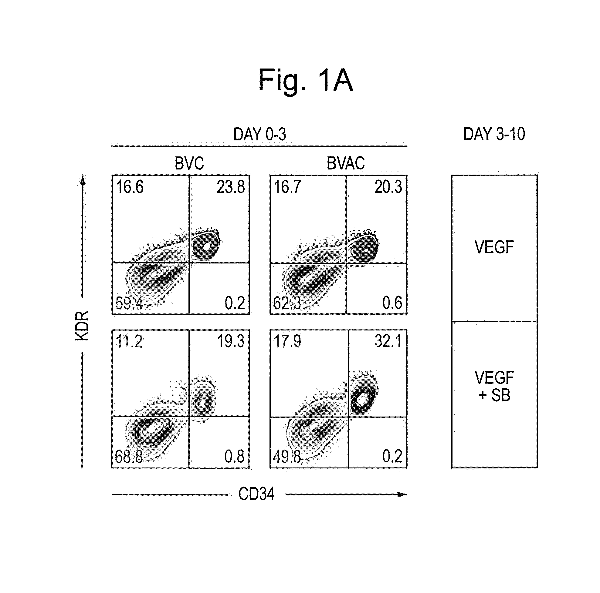
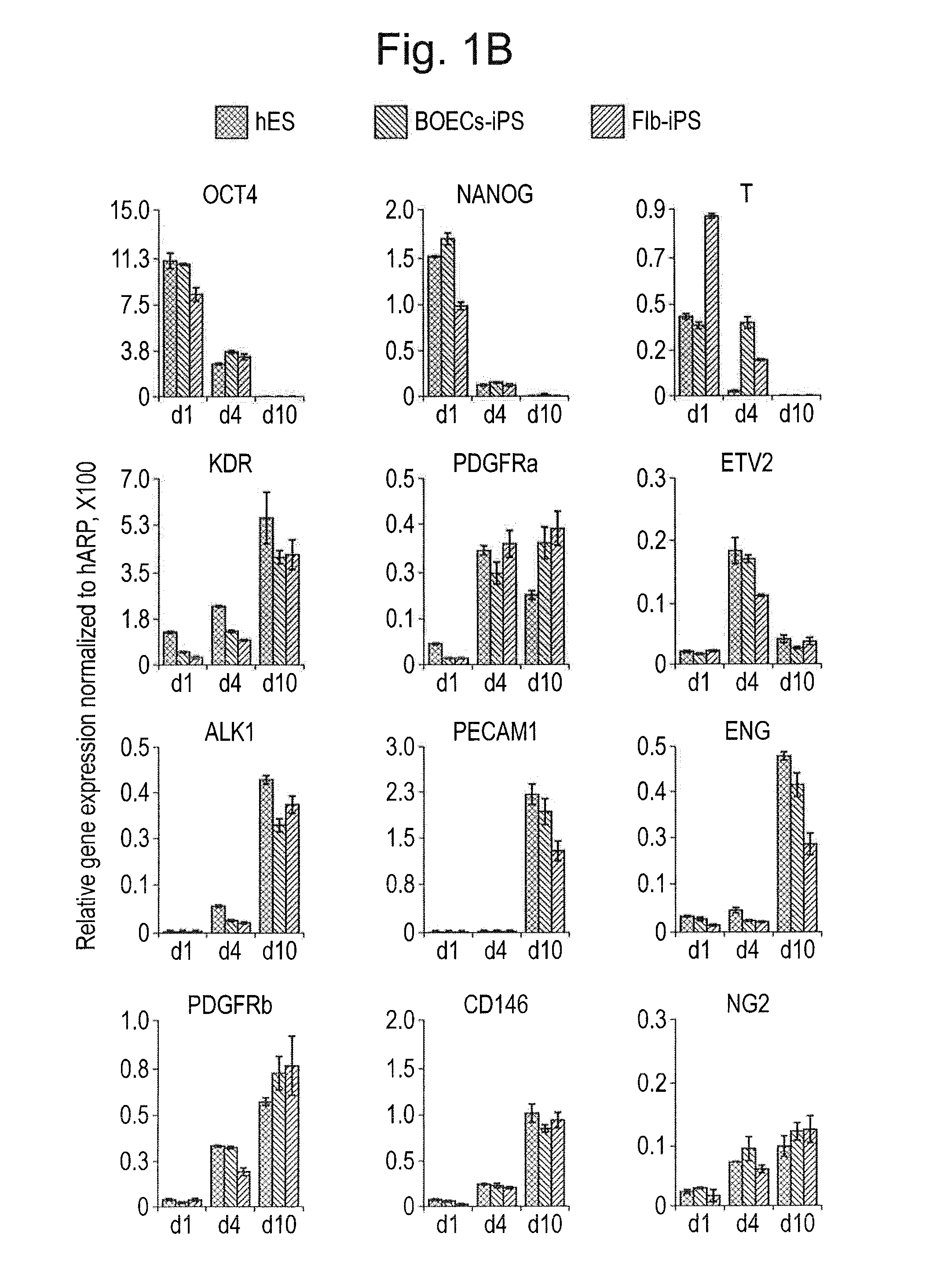
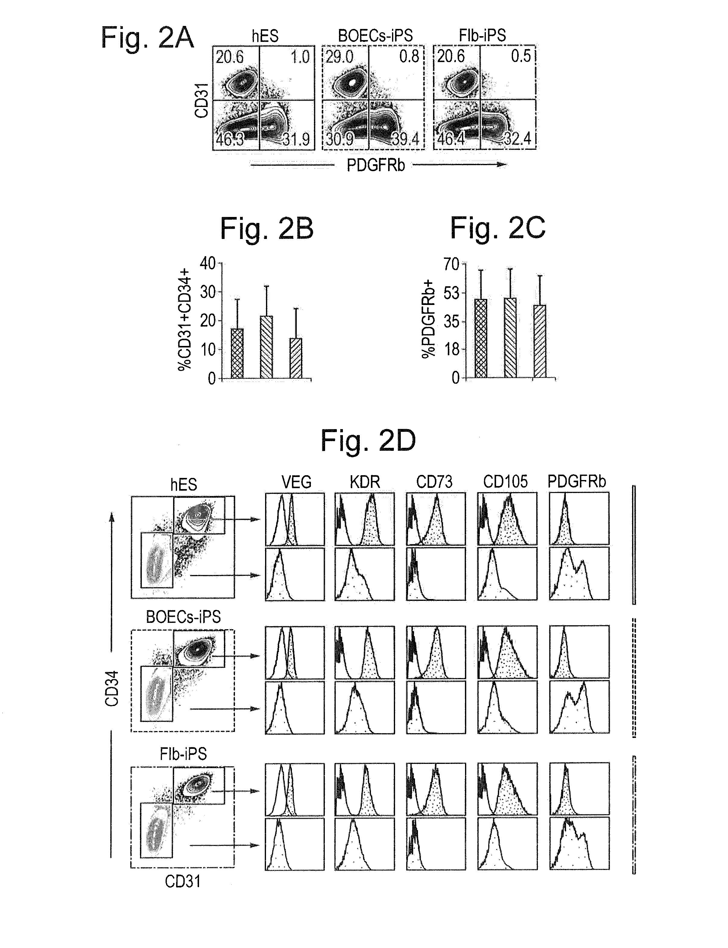
[0081]hiPSC lines were generated from Fib or BOECs using conditional lentiviral vectors containing the four transcription factors oct4, c-myc, Klf4 and Sox2, and characterized as described previously (Dambrot et al., 2013; Freund et al., 2008; Orlova et al., manuscript in preparation). hiPSCs and hESC (line HES-NL4; (van de Stolpe et al., 2005)) were routinely maintained on growth factor reduced MATRIGEL®-(BD, 354230) coated plates in mTeSR media (StemCell Technologies, 05850) and passaged mechanically. Differentiation was induced three days after passaging colonies by replacing mTeSR medium with the differentiation media based on BPEL (without PVA) (Ng et al., 2008) with timed addition of the following growth factors: 25 ng / ml ActivinA (Miltenyi, 130-095-547), 30 ng / ml BMP4 (Miltenyi, 130-095-549), VEGF165 (R&D Systems, 293-VE) and small molecule inhibitor CHIR99021 (Tocris, 4423). At day 3 and day 7 of differentiation, the media was refreshed...

example 2

Results

Derivation and Characterization of HHT1-iPSCs

[0110]Numerous iPSCs clones were generated from somatic tissue from two HHT1 patients with different ENG mutations (FIGS. 6A-6E) with normal efficiencies using standard reprogramming methods (Dambrot et al., 2013) indicating that the mutations did not affect reprogramming as such. Mutations were confirmed by sequencing of genomic DNA isolated from HHT1 patient-specific and control iPSC lines (FIG. 6B). Undifferentiated iPSC lines were maintained on MATRIGEL® in mTESR1 medium for at least 25 passages and expressed typical markers of pluripotent stem cells as shown by immunostaining (FIG. 6C). When allowed to differentiate spontaneously in culture all iPSC lines readily gave rise to the derivatives of the three germ layers, e.g., neurons expressing βIII-tubulin (ectoderm), endodermal cells expressing alphafetoprotein (AFP) and SMCs expressing smooth muscle actin (mesoderm) (FIG. 6E). To confirm pluripotency of the undifferentiated iP...


PUM

PropertyMeasurementUnit
temperatureaaaaaaaaaa
real time PCRaaaaaaaaaa

Abstract

The disclosure is concerned among others with means and methods for obtaining endothelial cells and to means and methods for in vitro cell culture comprising endothelial cells and pericytes and / or smooth muscle cells derived from the pericytes. The endothelial cells or the pericytes and / or smooth muscle cells, or both, are preferably derived from in vitro differentiated pluripotent stem cells.

Description

CROSS-REFERENCE TO RELATED APPLICATIONS[0001]This application is a national phase entry under 35 U.S.C. §371 of International Patent Application PCT / NL2014 / 050373, filed Jun. 10, 2014, designating the United States of America and published in English as International Patent Publication WO 2014 / 200340 A1 on Dec. 18, 2014, which claims the benefit under Article 8 of the Patent Cooperation Treaty and under 35 U.S.C. §119(e) to Dutch Patent Application Serial No. 2010949, filed Jun. 10, 2013.TECHNICAL FIELD[0002]The disclosure relates to the field of cell biology and medicine. The disclosure, in particular, relates to means and methods for in vitro production of endothelial cells, pericytes, pericyte-derived smooth muscle cells and vasculature. The disclosure further relates to means and methods for screening compounds for their angiogenic or anti-angiogenic activity. Produced endothelial cells can be cultured and cryopreserved.BACKGROUND[0003]Pluripotent stem cells (PSCs) proliferate o...

Claims


Application Information

Patent Timeline
Patent Type & Authority Applications(United States)
IPC IPC(8): C12N5/071G01N33/50
CPCC12N2501/155C12N5/0691G01N33/5061C12N5/069C12N2506/45C12N2501/15C12N2501/16C12N2501/165C12N2501/415G01N33/5064C12N2500/90C12N2501/727C12N2503/06
Inventor MUMMERY, CHRISTINE LINDSAYORLOVA, VALERIYA VIKTOROVNA
Owner ACADEMISCH ZIEKENHUIS BIJ DE UNIV VAN AMSTERDAM ACADEMISCH MEDISCH CENT
Learn More