Method for detection of fetal abnormalities

a technology for detecting abnormalities and fetal bodies, applied in the field of methods for detecting abnormalities of fetal bodies, can solve the problems of difficult medical conditions and decisions concerning abnormalities, common encounter of genetic diseases and chromosomal disorders, and significant risk

Inactive Publication Date: 2016-03-31
STELLING JAMES R
View PDF4 Cites 3 Cited by
  • Summary
  • Abstract
  • Description
  • Claims
  • Application Information

AI Technical Summary

Benefits of technology

The patent describes methods for performing a non-invasive prenatal test to evaluate fetal chromosomal and single gene disorders using trophoblast cells obtained from the cervix of pregnant women. The fetal mass is enriched in the sample, and the at least one trophoblast is isolated from the enriched sample. The genetic analysis can be performed using various genetic analysis techniques such as FISH, array comparative genomic hybridization (aCGH), karyomapping with single polymorphisms (SNPs), and next generation sequencing (Next Gen or NG). The technical effects include the non-invasiveness of the test and the accuracy of fetal genetic analysis for evaluating fetal chromosomal and single gene disorders.

Problems solved by technology

Genetic diseases and chromosomal disorders are unfortunately encountered commonly during pregnancies of many women.
These abnormalities often lead to very difficult medical conditions and decisions concerning these abnormalities.
Unfortunately, while these tests are highly accurate they do carry significant risk.
Therefore these invasive tests are usually limited to those patients considered to be at highest risk, based upon age and the mentioned biochemical and ultrasound markers.
Because of the risk and fear of these procedures many patients do not elect to undergo these procedures, potentially leading to the birth of more affected children.
This direction has been hampered by the low percentage of fetal cells found in maternal blood.
Cell free fetal DNA has also more recently been used but is less accurate then the invasive tests available.

Method used


Image

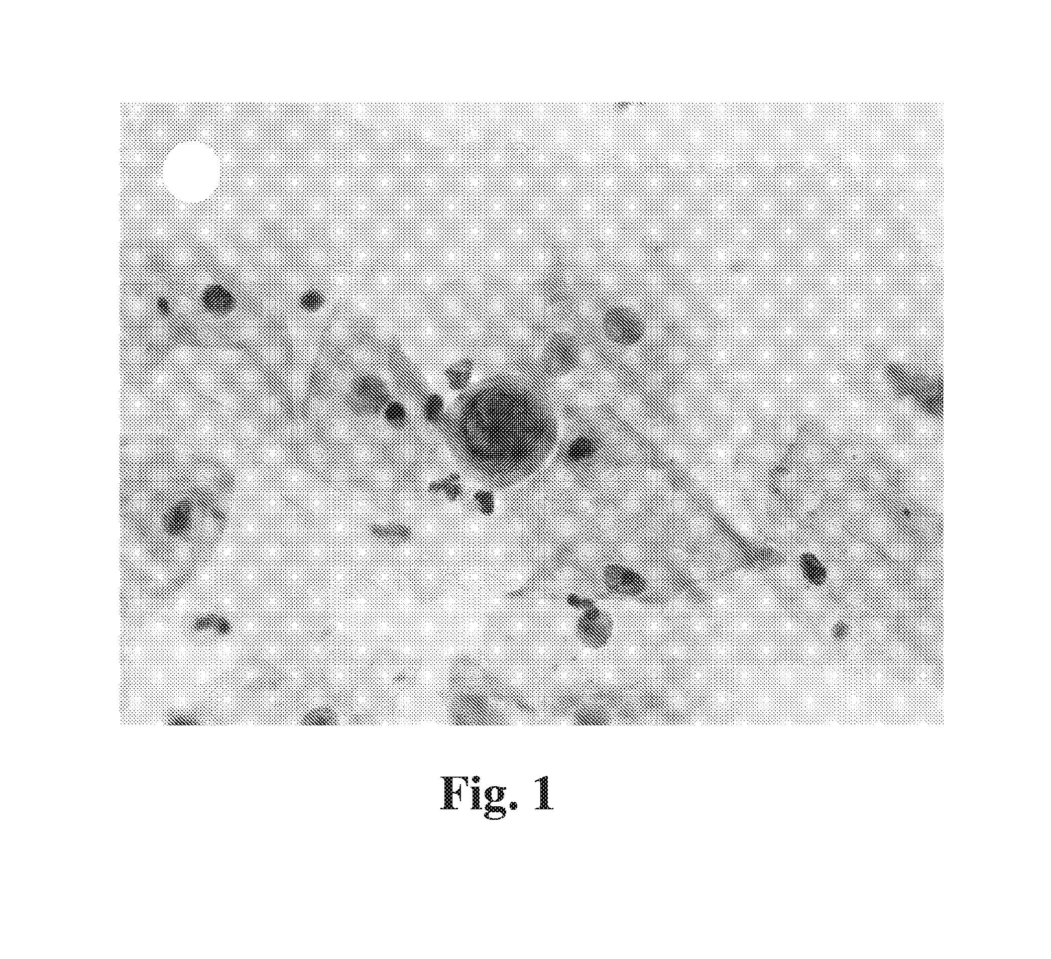
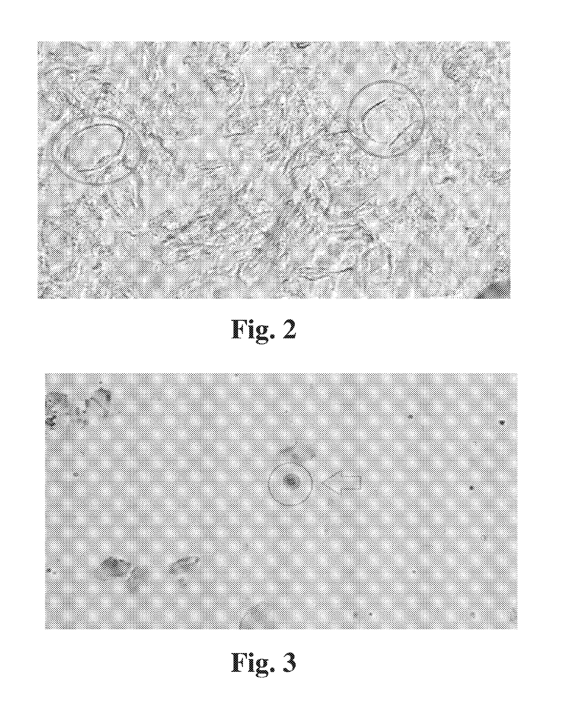
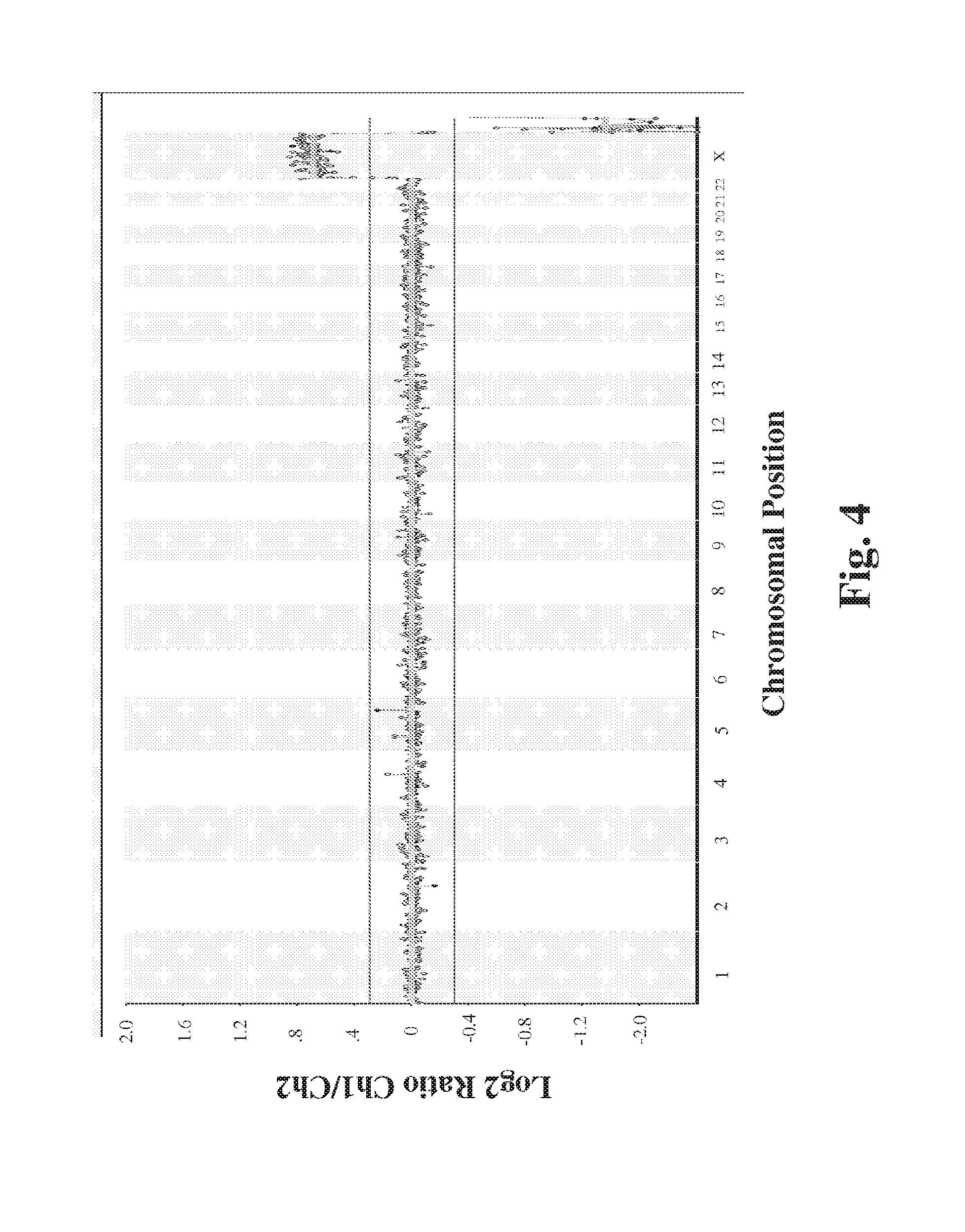
  • Method for detection of fetal abnormalities
  • Method for detection of fetal abnormalities
  • Method for detection of fetal abnormalities

Examples


example 1

Sample Collection

[0034]Cell sample from cervical swab. A Pap smear cytobrush is inserted through the external os to a maximum depth of 2 cm and removed while rotating it a full turn (i.e., 360°). In order to remove cells from the brush, the brush is swirled in a test tube or vial containing 2-3 ml of either a methanol-based solution such as THINPREP PRESERVCYT solution (Hologics, Inc.), or PBS with or without 10% serum albumin

example 2

Trophoblast Enrichment by Density Gradient Centrifugation

[0035]Cells in medium were enriched by applying the sample to a silane-coated silica particle density gradient (ALLGRAD, LifeGlobal Media, Inc.) First the sample was centrifuged in collection media to concentrate cellular material and remove any methanol. The pellet was isolated and placed in a prepared density gradient conical tube with 10-15 layer of varying dilutions of density gradient (ranging from 5%-70%). The tubes were centrifuged at 1200 g for 10-20 minutes. The fraction of sample between 30%-50% density gradient was isolated, resuspended in 5 mL PBS with or without 10% serum albumin, and centrifuged at 1000 g for 5 minutes. The pellet was then isolated and resuspended in 1 mL PBS with 10% serum albumin.

[0036]Surprisingly, the inventors found that density gradient centrifugation could successfully enrich trophoblasts in a sample obtained from the maternal cervix. Enrichment of trophoblasts was unexpected because recov...

example 3

Trophoblast Enrichment by Collagen Adhesion

[0037]As an alternative to density gradient centrifugation, collagen adhesion is utilized to enrich trophoblasts in a sample. A cervical cell sample is seeded onto GROWCOAT collagen-1 coated plates (Sarstedt, Inc., Newton, N.C.) and cultured according to the manufacturer's instructions. Trophoblasts adhere to the collagen surface and cervical cells and debris can be removed by washing. Following the washing step, the trophoblast-enriched sample can be removed by enzymatic digestion, such as trypsin or HYQTASE (HyClone, Inc.) for further isolation of trophoblasts.


PUM

PropertyMeasurementUnit
Adhesion strengthaaaaaaaaaa

Abstract

Disclosed are methods for non-invasive fetal genetic analysis involving enrichment of trophoblast cells in a maternal cervical sample, followed by isolation and genetic analysis of the isolated trophoblasts.

Description

CROSS-REFERENCE TO RELATED APPLICATIONS[0001]This application claims priority to U.S. provisional application 61 / 833,653, filed Jun. 11, 2013, which is incorporated herein in its entirety.BACKGROUND OF THE DISCLOSURE[0002]Genetic diseases and chromosomal disorders are unfortunately encountered commonly during pregnancies of many women. The rate of chromosomal abnormalities, such as Down Syndrome (Trisomy 21) increase with advancing maternal age. These abnormalities often lead to very difficult medical conditions and decisions concerning these abnormalities. Current obstetrical care involves several methods to help women predict the likelihood of chromosomal abnormalities during pregnancy. These techniques include some noninvasive tools such as ultrasound to measure nuchal thickness, as well as blood work such as the quad screen or triple screen. These noninvasive tests combined with patient's age will help predict the rate of having a chromosomal abnormal pregnancy. These tests will...

Claims


Application Information

Patent Timeline
IPC IPC(8): C12Q1/68
CPCC12Q1/6883C12Q2600/156C12Q2600/158G01N33/5091G01N2800/385
Inventor STELLING, JAMES, R.
Owner STELLING JAMES R
Why Eureka
Try Eureka