
Plasmonic substrate for multiplex assessment of type 1 diabetes

a plasmonic substrate and multiplex technology, applied in the field of nanostructured plasmonic metal films on substrates, can solve the problems of unsatisfactory diagnosis, no longer clear, up to 25% of children have unnecessary stress and health care expenditure, etc., and achieve the effect of detecting diabetes autoantibodies, rapid and sensitive diagnosis of diabetes, and equivalent sensitivity and specificity

Inactive Publication Date: 2016-01-28
THE BOARD OF TRUSTEES OF THE LELAND STANFORD JUNIOR UNIV +1
View PDF0 Cites 9 Cited by
  • Summary
  • Abstract
  • Description
  • Claims
  • Application Information

AI Technical Summary

Benefits of technology

The present invention is a new method for diagnosing diabetes by printing protein microarrays on a gold plasmonic substrate and detecting autoantibodies from a single patient sample. This approach allows for multiplexed testing for various autoimmune diseases, including celiac disease, autoimmune hypothyroidism, and Addison disease. The platform has been tested on children with new-onset diabetes and has shown similar sensitivity and specificity to traditional methods. The plasmonic gold chip platform has also been shown to have low background and a dynamic range that improves sensitivity in T1D diagnosis. The platform can detect both isotype-specific and total autoantibodies, and can be used as a research tool to test for putative novel biomarkers. The technology can also be used for point-of-care diagnosis and can help predict the efficacy of therapies and the natural history of diabetes development.

Problems solved by technology

This concurrent rise has resulted in a worldwide diagnostic dilemma and it is no longer clear what type of diabetes a symptomatic child has at disease presentation.
As a result, up to 25% of children have unnecessary stress and health care expenditure.
Unfortunately, this testing is cumbersome, is not available in resource-poor settings, and results are not available until weeks after a treatment plan has been selected.
Although RIA provides sensitive detection of diabetes autoantibodies, it requires radioisotopes, is time intensive, is expensive ($300-400), does not allow antigen multiplexing, and requires venipuncture.
Additionally, the Diabetes Autoantibody Standardization Program (DASP) workshops have reported unfavorable reviews of non-central laboratory RIA kits.
The current standard is performed with a radioimmunoassay, is very costly, cannot be multiplexed, is very expensive, and is not possible to perform in primary care and ER settings where it would be most effective and clinically useful.
More traditional and cheaper methods for detecting autoantibodies, including ELISA, have been unsuccessful.

Method used


Image

  • Plasmonic substrate for multiplex assessment of type 1 diabetes
  • Plasmonic substrate for multiplex assessment of type 1 diabetes
  • Plasmonic substrate for multiplex assessment of type 1 diabetes

Examples


example 1

Plasmonically Active Au Substrates

[0068]Plasmonic nanostructured gold substrates are fabricated, and are capable of optimal fluorescence enhancement up to 1000 fold for NIR fluorophores, as described in the above-referenced patent application and publication by Tabakman et al. Chemical synthesis and lithographic patterning techniques are used for the formation of plasmonic gold films on glass capable of maximizing the fluorescence enhancement of NIR dyes coupled to antibodies to the diabetes autoantigens in a patient sample. The plasmonic resonance is controlled by the gold nanostructure patterns to match to emission of NIR fluorophores. Nano-gaps between gold nanostructures are tuned to maximize local electrical field enhancement for optimal NIR-FE for fluorophores such as cy5, cy5.5, Atto 647N, cy7, and IR800.

example 2

Detection of Diagnostic Biomarkers

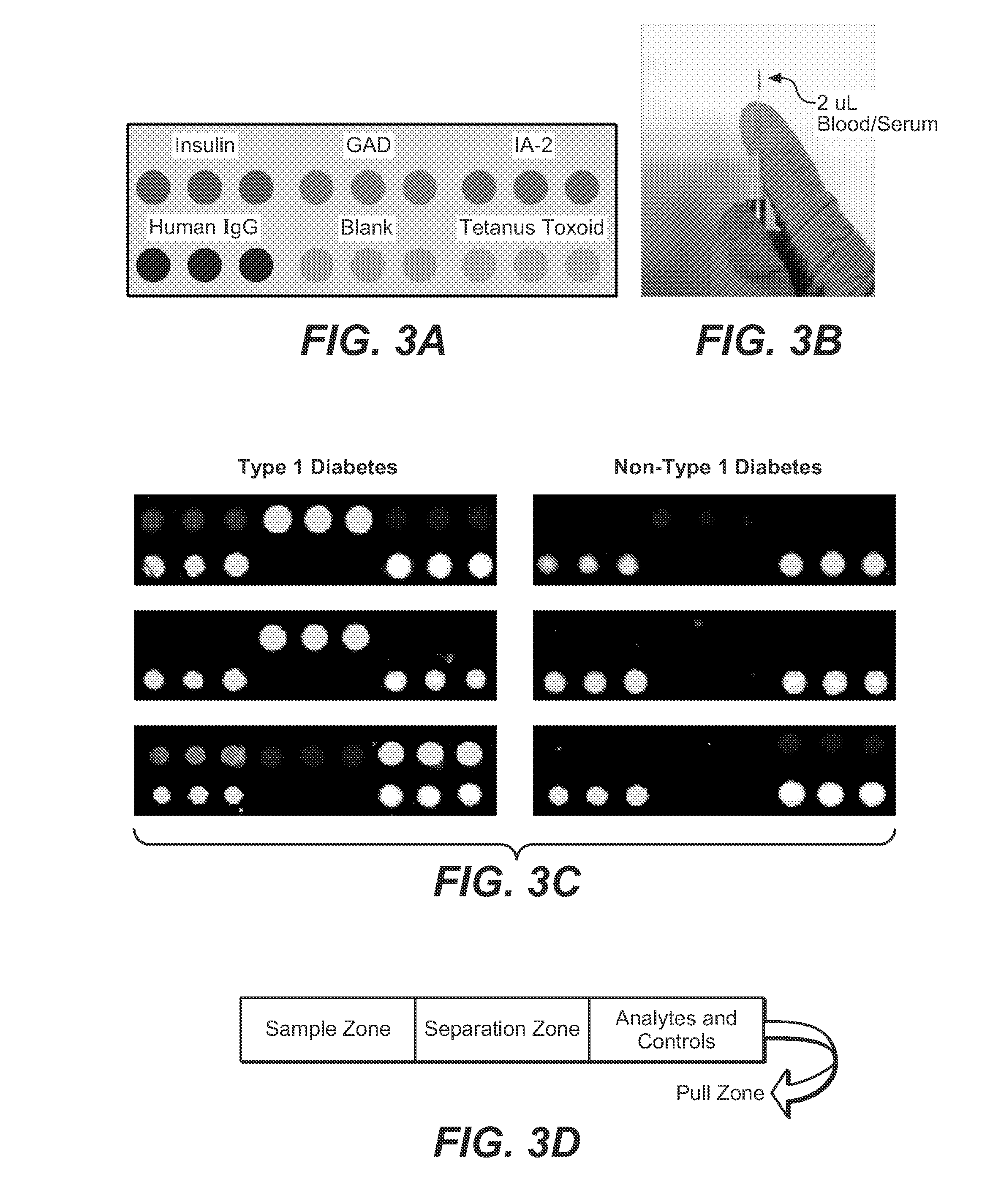
[0069](Taken from copending Ser. No. 13 / 728,798). This example employs protein microarray printing onto a gold plasmonic substrate on a glass chip as described in the examples above. This array allows detection of autoantibodies in a patient sample. The antibodies associated with diabetes type I are raised against small volumes of antigen. The present array and use of the plasmonic substrate with enhanced near-infrared fluorescence provides significantly improved sensitivity, as the gold film amplifies the fluorescent signal labels on detection antibodies. Additionally, this platform allows for multiplexed testing for more than one diabetes autoantibody from a single patient sample, in addition to testing for diagnostic autoantibodies for other autoimmune diseases known to have increased prevalence in patients with type 1 diabetes from the same sample (including celiac disease, autoimmune hypothyroidism, and Addison disease).

example 3

Surface Chemistry for Applying Antigens for Effective Antibody Capture

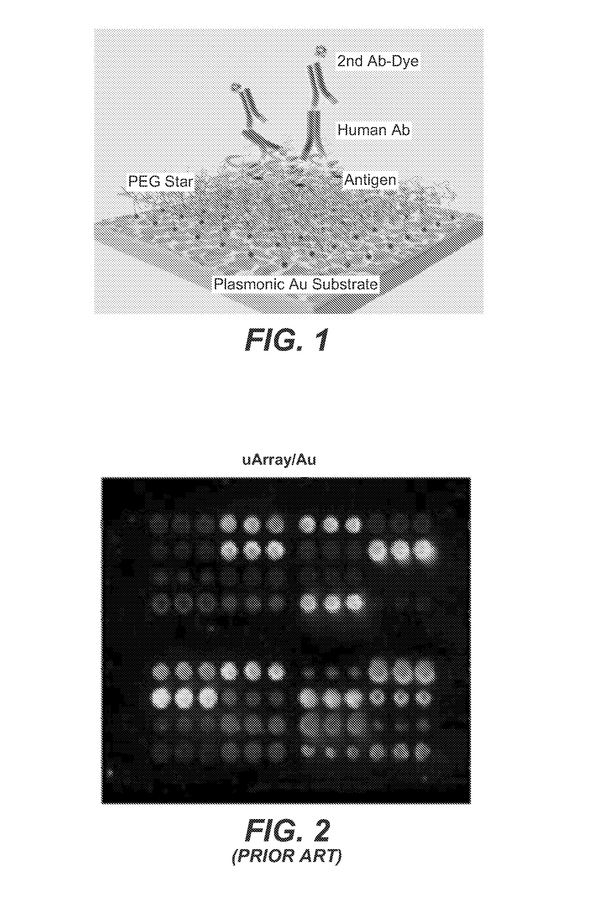
[0070]While fluorescence-enhancement will enhance positive binding signal, effective surface chemistry on Au plasmonic substrates is used herein to immobilize capturing antibodies and prevent non-specific binding, thus optimizing positive binding and reducing background and false signal. We here modify the plasmonic gold film for immobilization of multiplexed protein probes by first making self-assembled monolayers (SAM) of thiol-containing molecules terminated with carboxylate groups. Branched hydrophilic polymers of 6-arm-poly(ethylene glycol) (PEG)-NH2 is then grafted to SAMs, followed by reaction with succinic anhydride to obtain carboxylic acid groups off the 6-arm-PEG for protein immobilization. This method is surprisingly advantageous, since it employs branched-PEG star polymers as a hydrophilic, biocompatible cushion (FIG. 1) on Au to allow oriented immobilization of protein probes with minimal conformati...


PUM


Abstract

Disclosed are methods and materials providing fluorescence detection of autoantibodies present in individuals who have developed or are at risk for type 1 diabetes. Provided is a plasmonic chip capable of fluorescence-enhancement of >100-fold. The fluorescent signal is generated by an anti-human antibody antibody, such as an anti-IgG antibody that is coupled to a fluorophore selected to emit at a wavelength enhanced by the plasmonic chip, for example in the NIR.

Description

CROSS-REFERENCE TO RELATED APPLICATIONS[0001]This application claims priority from U.S. Provisional Patent Application No. 61 / 783,605, filed Mar. 14, 2013, and U.S. Provisional Patent Application No. 61 / 911,315, filed Dec. 3, 2013, which are hereby incorporated by reference in their entirety.STATEMENT OF GOVERNMENTAL SUPPORT[0002]This invention was made with Government support under contract TW008781 and contract CA135109 awarded by the National Institutes of Health. The Government has certain rights in this invention.REFERENCE TO SEQUENCE LISTING, COMPUTER PROGRAM, OR COMPACT DISK[0003]None.BACKGROUND OF THE INVENTION[0004]1. Field of the Invention[0005]The present invention relates to the formation of nanostructured plasmonic metal films on substrates, where such plasmonic films are useful for spectroscopy and immunoassays, and, in particular to a plasmonic substrate used for detection of autoantibodies indicative of type I diabetes.[0006]2. Related Art[0007]Presented below is bac...

Claims


Application Information

Patent Timeline
IPC IPC(8): G01N33/68
CPCG01N2800/042G01N33/6854G01N33/54373
Inventor FELDMAN, BRIAN JAYDAI, HONGJIEKUMAR, RAJIV B.ZHANG, BO
Owner THE BOARD OF TRUSTEES OF THE LELAND STANFORD JUNIOR UNIV
Learn More