Pericardial heart valve replacement and methods of constructing the same

a technology of pericardial valves, which are applied in the field of constructing a pericardial heart valve replacement, can solve the problems of more prone to damage of replacements

Inactive Publication Date: 2015-11-26
STRAKA FRANTISEK +1
View PDF3 Cites 4 Cited by
  • Summary
  • Abstract
  • Description
  • Claims
  • Application Information

AI Technical Summary

Benefits of technology

The patent describes a method for creating a heart valve replacement using a patient's own tissue. An anatomically tailored stent is created to match the patient's native heart valve and the blood vessels near it. The patient's pericardial tissue is then sutured to the stent. This method makes it easier for the new heart valve to settle in its proper position and function properly. The use of autologous tissue also reduces the risk of rejection or complications. Overall, this method creates a heart valve replacement that is safe and effective.

Problems solved by technology

In the case that three pieces of pericardium are used to suture the heart valve replacement it would be necessary to suture the individual pieces at the commissures which would make the replacement more prone to damage.

Method used


Image

  • Pericardial heart valve replacement and methods of constructing the same
  • Pericardial heart valve replacement and methods of constructing the same
  • Pericardial heart valve replacement and methods of constructing the same

Examples


first embodiment

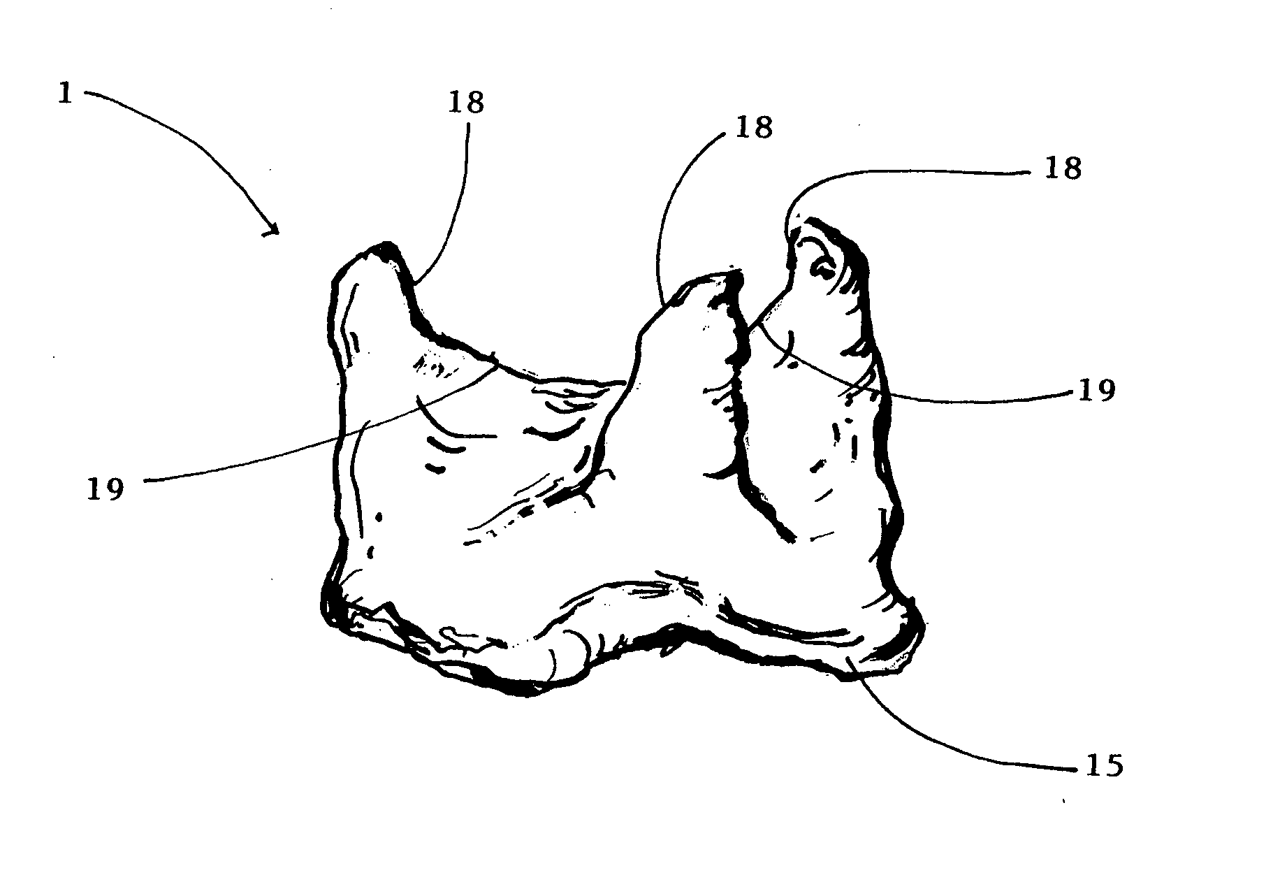
[0048]The stents 2 for construction of a living human pericardial heart valve replacement 1 are designed in two embodiments. In the first embodiment the sewing cuff 15 is attached to the stent 2 and is a part of the pericardium from which the heart valve replacement 1 is sutured.

second embodiment

[0049]In the second embodiment the stent 2 and the sewing cuff 15 are two separate components assembled together with sutures or clips after the pericardium has been sutured to the stent 2. This separate arrangement simplifies suturing the pericardium to the stent 2 and allows for the use of larger sewing cuffs 15 when irregularities in the aortic annulus need be sealed. Stents 2 and sewing cuffs 15 are preferably coated or impregnated with various agents to enhance their resistance to thrombosis, anticalcification or retard pannus overgrowth, such as alpha oleic acid or Carbofilm.

[0050]The stentless living pericaridial heart valve replacement 1 for open surgical implantation involves suturing the human pericardial heart valve replacement 1 directly in place of the native heart valve without the use of a stent 2. The size and shape of the heart valve replacement 1 annulus and height of commissural attachments is determined by three-dimensional imaging (echocardiography, CT or MRI) o...


PUM


Abstract

The pericardial heart valve replacement (1) is constructed from living tissue of autologous pericardium containing living pericardial interstitial cells (PICs) and a living extracellular matrix (ECM) in the following way: in the first step the size and shape of the patient's native pathological heart valve is determined using real-time three-dimensional transesophageal echocardiography (TEE) or computer tomography (CT angiography) or magnetic resonance imaging (MRI); in the next step a stented (2) or a stentless pericardial heart valve replacement (1) is constructed from a single sheet of living autologous pericardium obtained from the patient; the size and shape of the pericardial heart valve replacement (1) corresponds to the size and shape of the patient's native heart valve and aortic or pulmonary root. The subject matter of the invention also includes the development of a device (5) for the conditioning and modification of the living autologous pericardial heart valve replacement (1) consisting of a ipulsatile pump (6) driving the flow of culture medium in the device (5) to perfuse the cells in the pericardium, a reservoir (7) regulating the fluid capacitance and function of the pericardial heart valve replacement (1), a chamber (8) containing a holder (9) for the placement of the pericardial heart valve replacement (1) during dynamic conditioning, and a gas exchanger (10a, b) for perfusing C02 into the culture medium. The reservoir (7), the chamber (8) and the gas exchanger (10a, b) are placed in an incubator (11) with an inner atmosphere of air mixed with 5% C02 and a temperature of 36 to 37° C., the individual components of the device (5) being connected by tubing (12).

Description

FIELD OF THE INVENTION[0001]The present invention relates to a method of constructing a pericardial heart valve replacement, a pericardial heart valve replacement constructed according to this method, and a device for the in vitro conditioning and modification of tissue of autologous pericardium for use as a pericardial heart valve replacement.BACKGROUND OF THE INVENTION[0002]The implantation of heart valve replacements is an increasingly frequent medical procedure. One of the principal topics of cardiac surgery and cardiology is the construction of heart valve replacements for clinical use.[0003]Presently, whole porcine valves are commonly used as heart valve replacements in humans or the cusps of heart valve replacements are constructed from bovine pericardium. The disadvantage of such heart valve replacements is that they are xenogeneic to the immune system of the human patient and thus immunogenic. The patient's immune system reacts to such heart valve replacements and induces t...

Claims


Application Information

Patent Timeline
IPC IPC(8): A61F2/24
CPCA61F2/2412A61F2240/001A61F2/2418A61F2/2415A61L27/3604A61L27/3691A61F2/2472A61F2240/002A61L27/507
Inventor STRAKA, FRANTISEKMAS N, JAROSLAVSCHORN K, DAVIDPIRK, JAN
Owner STRAKA FRANTISEK
Why Eureka
Try Eureka