
Simulation-based estimation of elasticity parameters and use of same for non-invasive cancer detection and cancer staging

Active Publication Date: 2015-08-06
THE UNIV OF NORTH CAROLINA AT CHAPEL HILL
View PDF0 Cites 8 Cited by
  • Summary
  • Abstract
  • Description
  • Claims
  • Application Information

AI Technical Summary

Benefits of technology

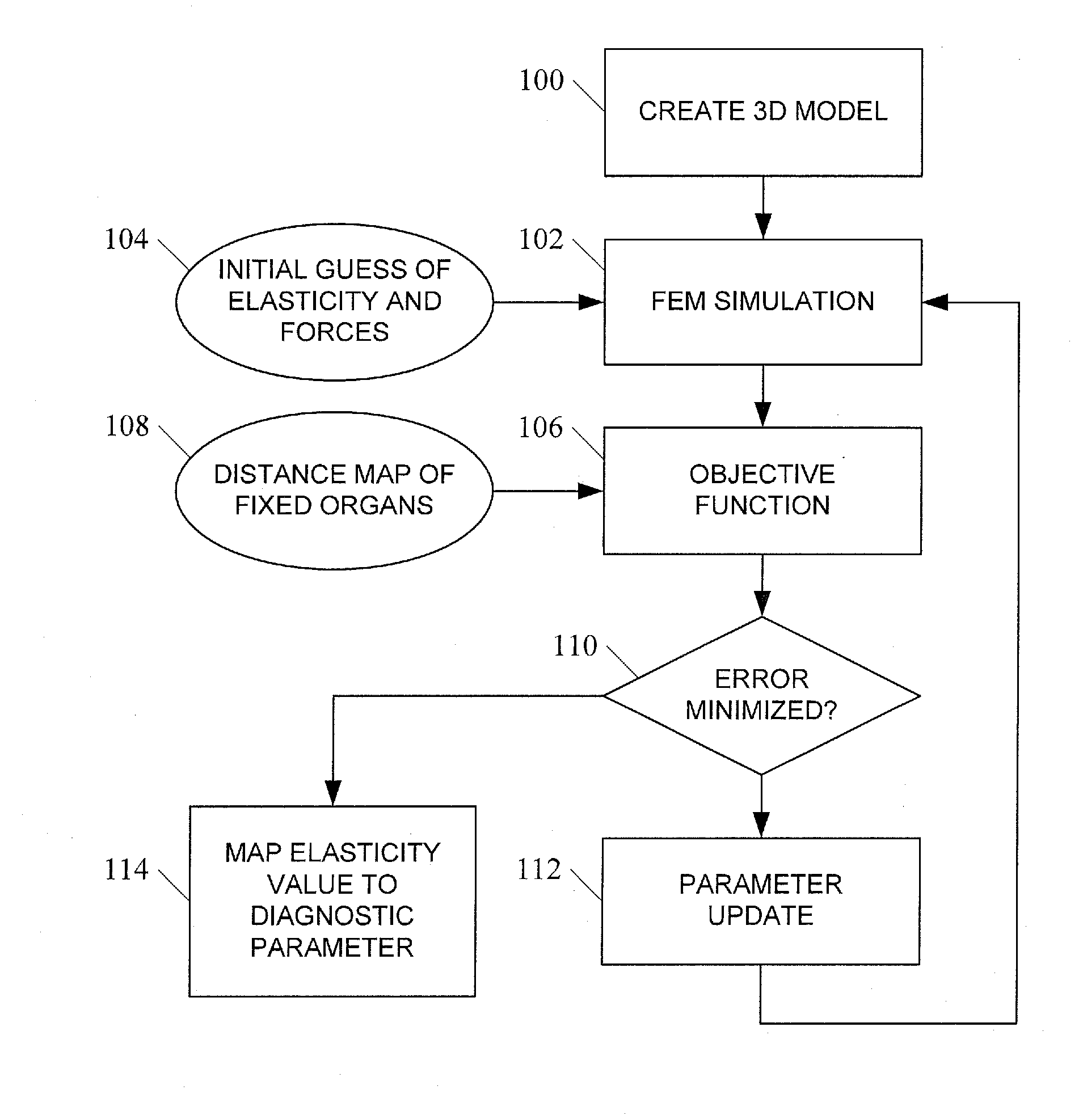
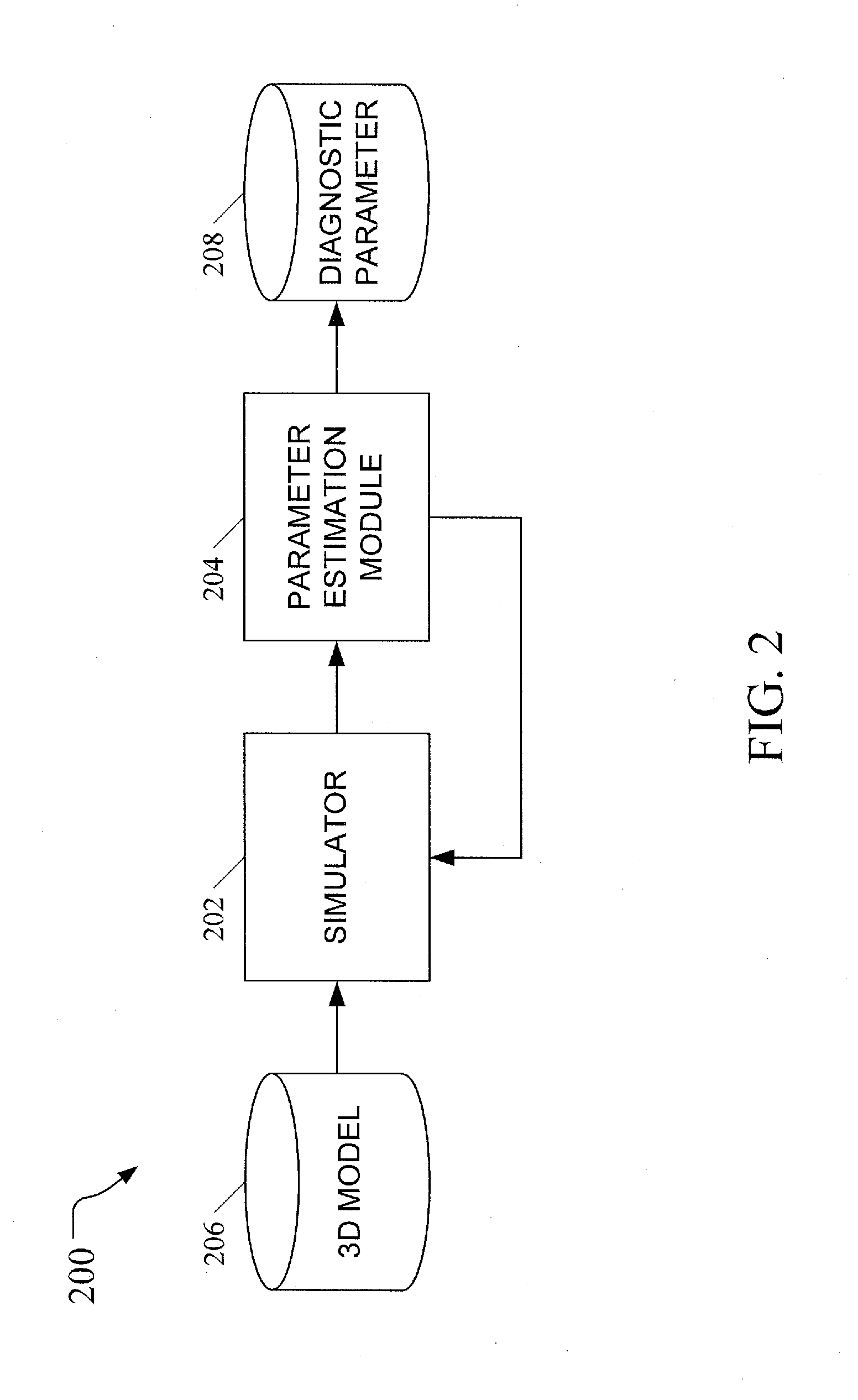
The patent describes a method and system for automatically estimating elasticity and boundary forces in a 3D model of biological tissue. This is done by simulating the application of an external force and measuring the resulting deformation of the model. The elasticity value is then mapped to a diagnostic parameter, such as an indication of the possibility, presence, or stage of cancer. The technical effects of the patent include improved accuracy and efficiency in estimating elasticity parameters, as well as a non-rigid image registration approach that guarantees quality of deformation fields.

Problems solved by technology

Since we do not assume a good correspondence for pixels inside an object, the resolution of the resulting elastogram is limited to the object boundaries.
Moreover, due to the patient-to-patient differences, these material properties sometimes need hand adjustments.
Therefore the elasticity cannot be recovered.
These results suggest that our ability to recover the parameters is limited by how stiff the object is.
When an object has a very high stiffness, its shape becomes insensitive to the parameters.
In this case, the shape can still be recovered, but the resulting parameters may not be accurate.
Notice that the problem of solving for elasticity and for boundary forces is ill-posed with a single object.
A modified Newton's method has been used in elasticity reconstruction [14], but the Hessian matrices can only be approximated and are usually ill-conditioned.
Therefore we expect to see worse accuracy in the case of stiffer cancerous tissues.
Notice that in the elasticity recovery process, we do not know the extent of the tumor due to the imaging limitation, and we only recover one value for the prostate.
Our method enforces physically-based constraints and results in errors within the diameter of the spot.
However, the recovered values are less consistent than those from the linear FEM implementation.
While the performance is acceptable for simple models with low degrees of freedom, it may take more than eight hours for some complex 3D models with many thousands of degrees of freedom for boundary forces.
However, the generation of the example displacements is nontrivial and depends on the specifics of the application.

Method used


Image

  • Simulation-based estimation of elasticity parameters and use of same for non-invasive cancer detection and cancer staging
  • Simulation-based estimation of elasticity parameters and use of same for non-invasive cancer detection and cancer staging
  • Simulation-based estimation of elasticity parameters and use of same for non-invasive cancer detection and cancer staging

Examples


Embodiment Construction

[0029]In accordance with the subject matter disclosed herein, systems, methods, and computer readable media for simulation-based estimation of elasticity parameters and use of same for non-invasive cancer detection and cancer staging are provided. Reference will now be made in detail to exemplary embodiments of the present invention, examples of which are illustrated in the accompanying drawings. Wherever possible, the same reference numbers will be used throughout the drawings to refer to the same or like parts.

I. INTRODUCTION

[0030]We propose an entirely passive analysis of a pair of images that only uses information about the boundaries of corresponding internal objects. We assume the images have already been segmented, that is, the organ boundaries have been found. Since we do not assume a good correspondence for pixels inside an object, the resolution of the resulting elastogram is limited to the object boundaries. Namely, we assume that the elasticity is fixed within each objec...


PUM


Abstract

Simulation-based estimation of elasticity parameters and use of same for non-invasive cancer detection and cancer staging are disclosed. According to one aspect, a method for simulation-based estimation of elasticity parameters includes constructing a 3D model of an object comprising biological tissue, the model having a first shape and an elasticity value. A simulation iteration is then performed, which includes simulating the application of an external force to the model, causing the model to have a second shape, measuring the difference between the second shape and a target shape, and determining whether the measured difference between the second shape and the target shape is within a threshold of error. If the measured difference is not within the threshold of error, the process performs additional iterations using adjusted elasticity or force values until the measured difference is within the threshold of error, at which time the final elasticity and force values are reported. In one embodiment, the elasticity value is mapped to a diagnostic parameter, such as an indication of the possibility, presence, or stage of cancer.

Description

PRIORITY CLAIM[0001]This application claims the benefit of U.S. Provisional Patent Application Ser. No. 61 / 680,116, filed Aug. 6, 2012; the disclosure of which is incorporated herein by reference in its entirety.GOVERNMENT INTEREST[0002]This invention was made with government support under Grant No. W911NF-06-1-0355 awarded by the U.S. Army Research Office. The U.S. Government has certain rights in the invention.TECHNICAL FIELD[0003]The subject matter described herein relates to methods and systems for characterization of biological tissue elasticity using non-invasive means. More specifically, the subject matter described herein relates to simulation-based estimation of elasticity parameters, which may be used for non-invasive cancer detection and cancer staging.BACKGROUND[0004]Estimation of tissue stiffness is an important means of noninvasive cancer detection. Existing elasticity reconstruction methods usually depend on a dense displacement field (given by ultrasound or MR images...

Claims


Application Information

Patent Timeline
IPC IPC(8): A61B6/00G06T19/20G06T15/00A61B6/03
CPCG06T7/0012G06T7/0032G06T2207/10081G06T2207/30081G06T19/20G06T15/00G06T2219/2021A61B6/032A61B6/466A61B6/50A61B6/5217G06T2210/41G06T7/344
Inventor LEE, HUAI-PINGFOSKEY, MARK STEPHENNIETHAMMER, MARCLIN, MING
Owner THE UNIV OF NORTH CAROLINA AT CHAPEL HILL
Learn More