Method for diagnosing skin disease based on in vivo skin imaging
a skin disease and in vivo technology, applied in the field of human skin disease diagnosis, can solve the problems of not disclosing the use of the microscopic system as a device, harmful x-ray imaging methods, emotional distress of patients,
- Summary
- Abstract
- Description
- Claims
- Application Information
AI Technical Summary
Benefits of technology
Problems solved by technology
Method used
Image
Examples
example 1
Diagnosis of Melanoma
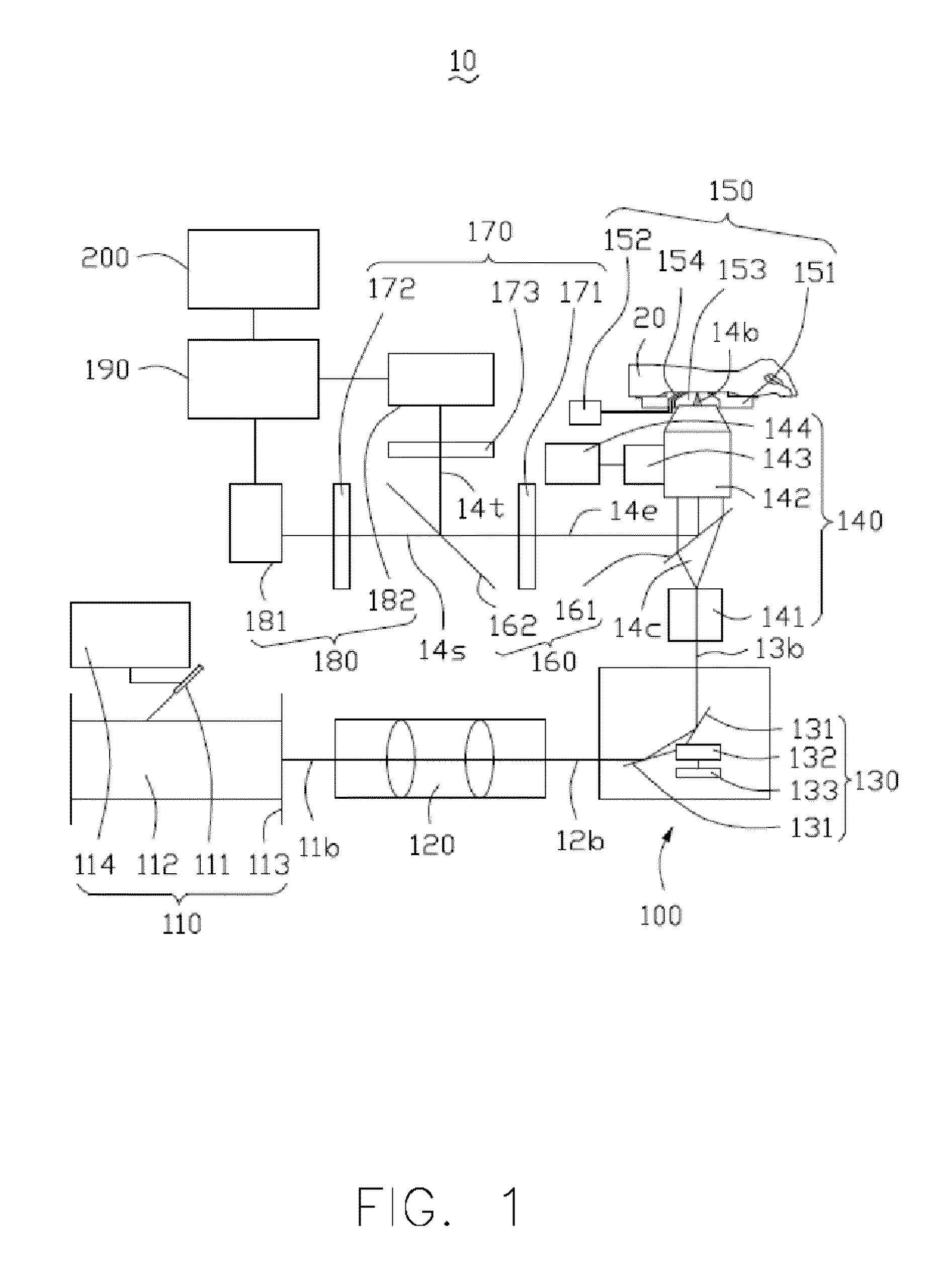
[0109]Melanoma is the most serious type of skin cancer, which is caused by malignant changes in melanocytes. Sectioned images of the skin tissue of a patient are obtained by the HGM device 100, and taken at different depths relative to the skin surface (10, 40, 65, 80, 110, and 150 μm) (see FIG. 15). Morphological features, including distribution of melanocytes, size of melanocytes, shape of melanocytes, size of melanocytic nuclei, shape of melanocytic nuclei, intracellular distance of melanocytes, and proliferation of melanocytes, are identified and analyzed through processing the sectioned image. Irregularly shaped cells with strong THG signals are found in the stratum granulosum, referring to arrows in FIGS. 15(a)-(b). The distribution of these cells is similar to the pagetoid spread of melanocytes observed in the conventional pathologic sections of melanoma. Deeper into the skin lesion, the proliferation of polymorphous THG-bright cells in the epidermis (see...
example 2
Diagnosis of Basal Cell Carcinoma
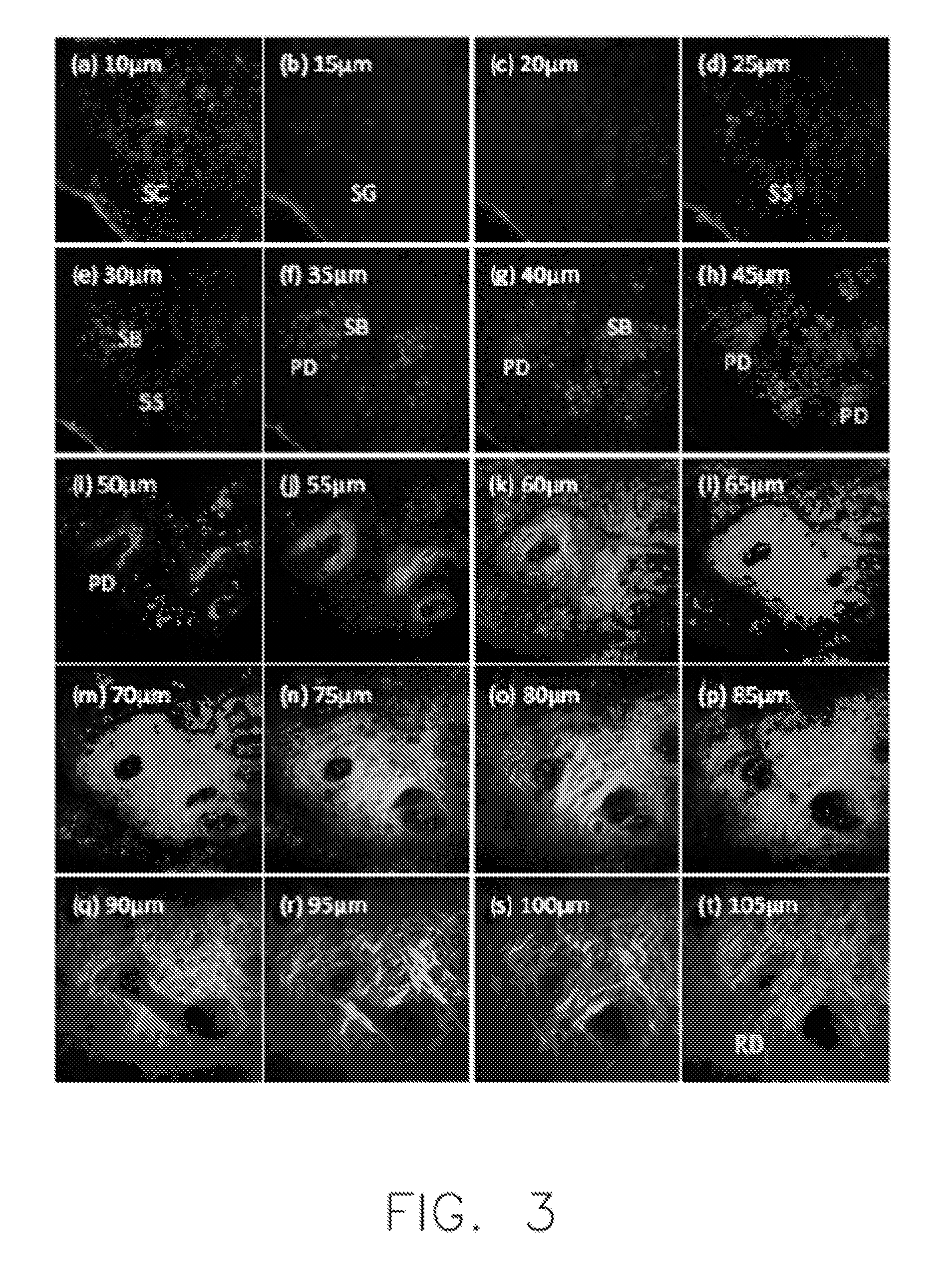
[0110]Basal cell carcinoma (BCC) is the most common type (>80%) of skin cancer, which grows from the basal cell layer. Sectioned images of the skin tissue of a patient are obtained by the HGM device 100, and taken at different depths relative to the skin surface (5, 15, 25, 45, 65, and 85 μm) (see FIG. 16). In FIGS. 16(a)-(c), “SC” stands for stratum corneum; “SG” stands for stratum granulosum; and “SS” stands for stratum spinosum. Morphological features, including shape of basal cells, size of basal cells, distribution of peripheral palisading cells, collagen bundles, dendritic processes of cells, and proliferation of basal cells, are identified and analyzed through processing the sectioned image. It is found that THG-bright cells rising from the stratum basale and extending into the dermis appear polymorphous with variations in size and shape (see FIGS. 16(d)-(f)). Moreover, the presence of elongated, peripheral palisading cells in the tumor nodule...
example 3
Diagnosis of Melanocytic Nevus
[0111]Melanocytic nevus is a form of benign neoplasm that can be divided into three nevus types according to the locations of the nevus cells, which are derived from melanocyte. Sectioned images of the skin tissue of a patient suffered from melanocytic nevus are obtained by the HGM device 100 (see FIG. 17). Morphological features, including size of nevus cells, shape of nevus cells, and distribution of nevus cells, are identified and analyzed through processing the sectioned image. The presence of the aggregation of THG-bright cells in the dermo-epidermal junction (see dashed circle in FIG. 17(a)) and in the dermis (see arrow in FIG. 17(d)) are observed in junctional and intradermal melanocytic nevi, respectively. The nevus cells in both the dermo-epidermal junction and dermis from the HGM images of compound nevi are also observed. The cells in the nests, which are monomorphic in size and shape, are observed using THG imaging. The scattered melanocytes ...
PUM
 Abstract
Description
Claims
Application Information
 - R&D
- Intellectual Property
- Life Sciences
- Materials
- Tech Scout
- Unparalleled Data Quality
- Higher Quality Content
- 60% Fewer Hallucinations
Browse by: Latest US Patents, China's latest patents, Technical Efficacy Thesaurus, Application Domain, Technology Topic, Popular Technical Reports.
© 2025 PatSnap. All rights reserved.Legal|Privacy policy|Modern Slavery Act Transparency Statement|Sitemap|About US| Contact US: help@patsnap.com


