
Dual Balloon Biliary Stone Extraction Device

a biliary stone and balloon technology, applied in the field of medical devices, can solve the problems of difficult selective cannulation of the biliary radicle (cystic duct, left/right intrahepatic duct, etc.) in patients with pathology located in the cystic duct, gallbladder or branches of the intrahepatic duct, and stones can get jammed in the end of the bile duct, so as to facilitate the selective cannulation of th

Inactive Publication Date: 2015-06-04
THE JOHN HOPKINS UNIV SCHOOL OF MEDICINE
View PDF0 Cites 8 Cited by
  • Summary
  • Abstract
  • Description
  • Claims
  • Application Information

AI Technical Summary

Benefits of technology

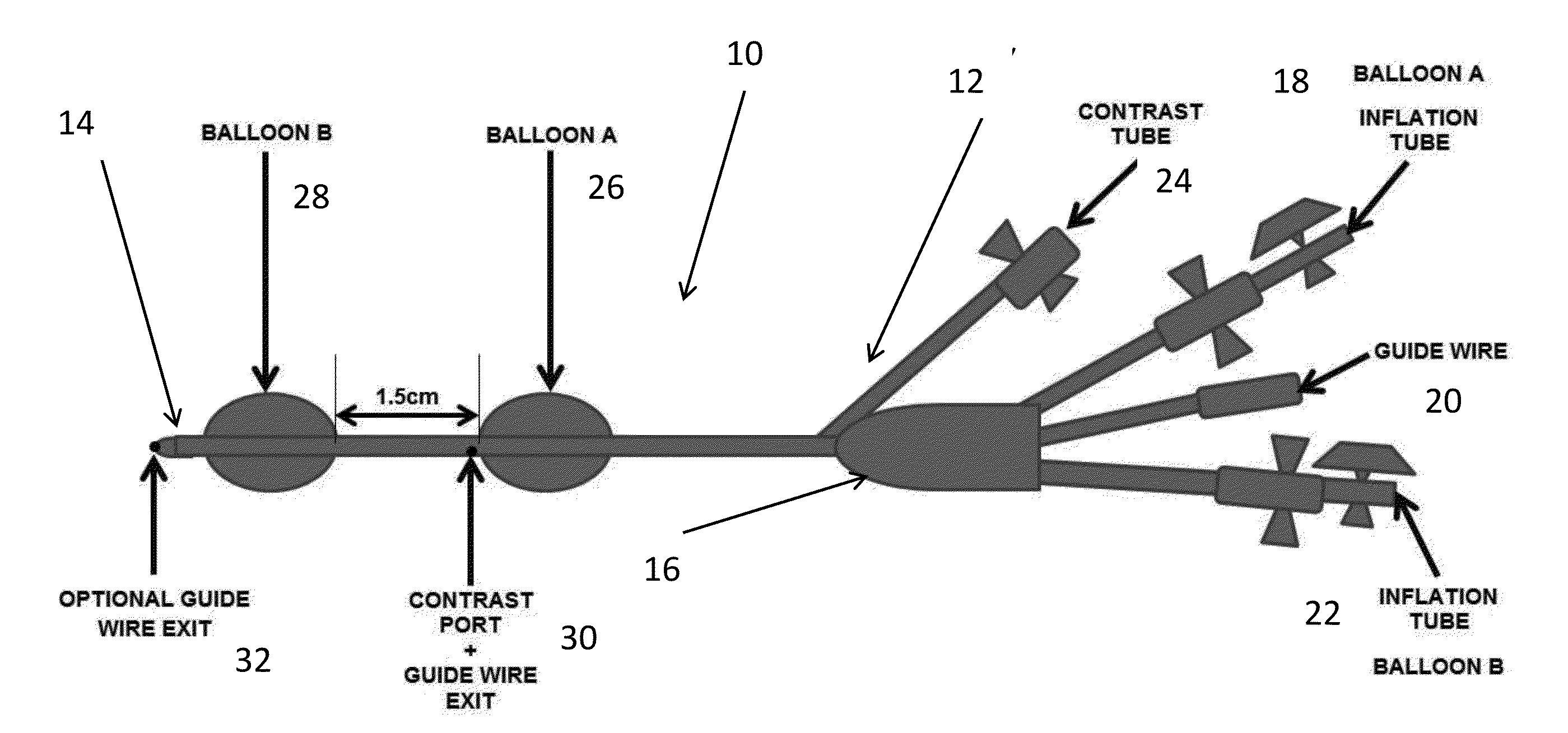
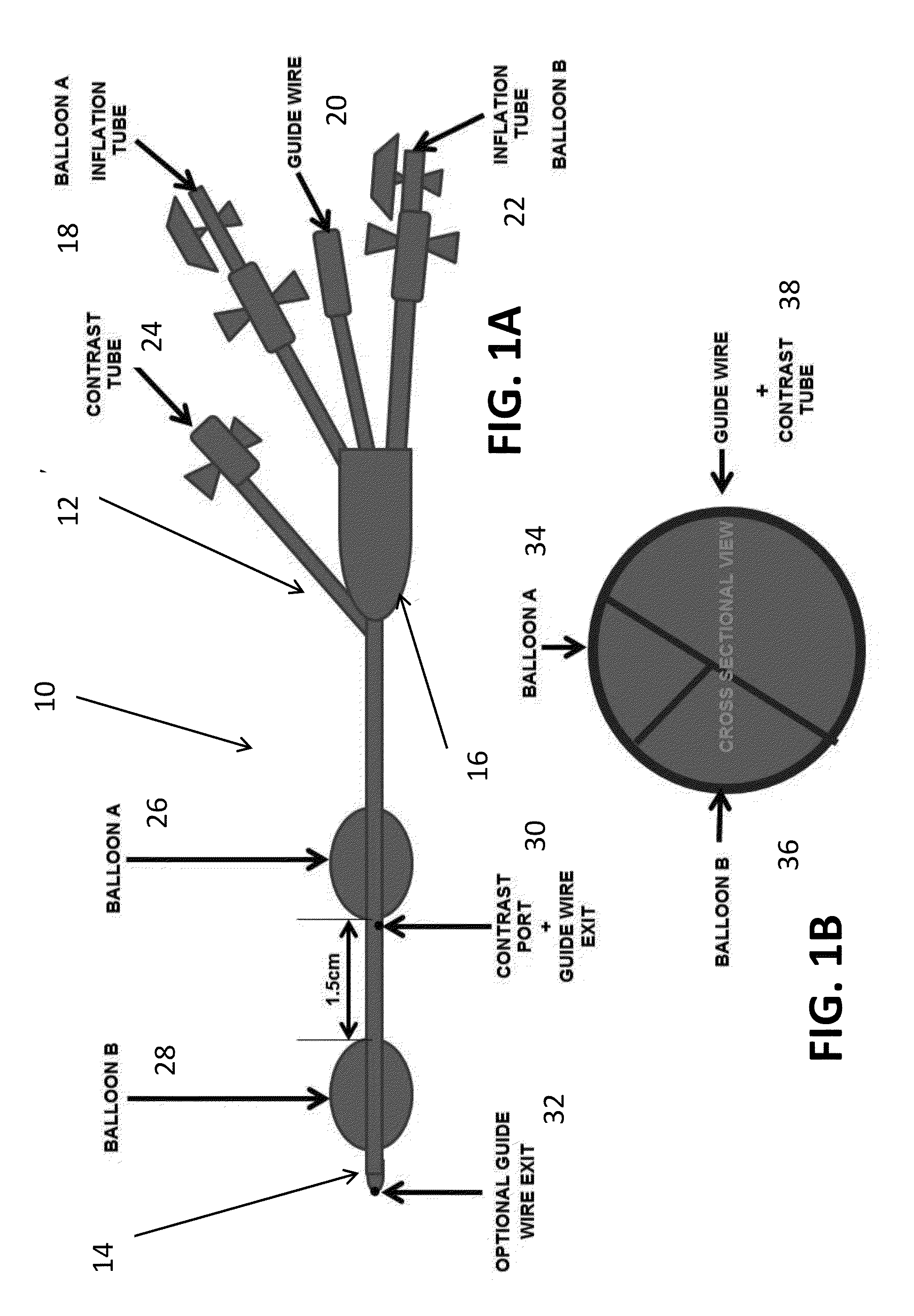
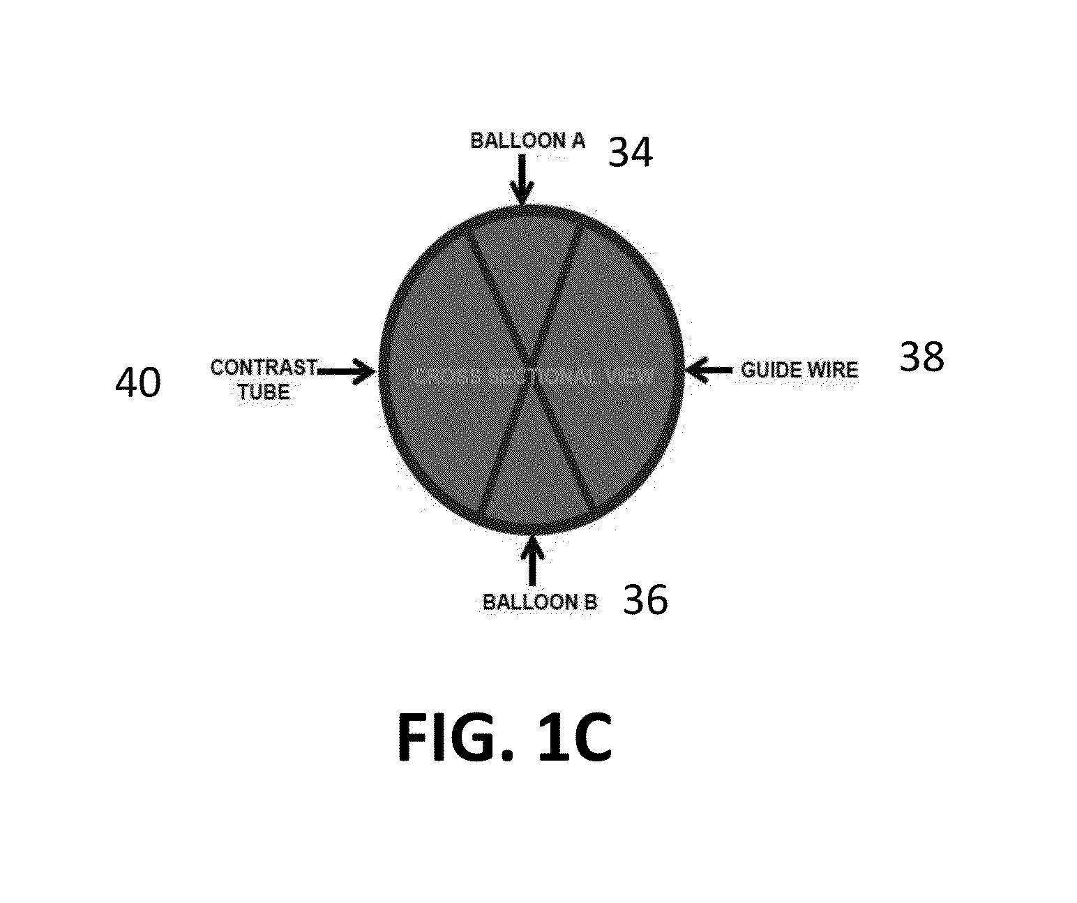
The present invention provides a method for removing calculi in the bile duct, which involves inserting two balloons into the duct and inflating them to capture the calculus. The catheter can then be withdrawn to allow the calculus to pass. The method also allows for hydrodynamic delivery of genes to treat biliary epithelium disease, and facilitates selective guide wire cannulation of a cystic duct or intrahepatic duct.

Problems solved by technology

Selective guide wire cannulation of a biliary radicle (cystic duct, left / right intrahepatic duct etc) in patients with pathology located in the cystic duct, gallbladder or branches of the intrahepatic ducts can also be difficult.
Guide wire placement in the cystic duct and intrahepatic ducts is often a challenge due to the state of current accessories (which only allow the guide wire to exit the catheter at its tip) and their narrow lumen (often 2 mm in diameter).
Biliary calculi and debris can become jammed in the distal common bile duct.
These stones can get jammed in the end of the bile duct due to a kink or a small biliary opening.
There is currently no large animal model for CCA.
Its delivery method has been limited to the vasculature though there does not appear to be a reason why the same delivery method cannot be used in other anatomical locations such as the bile duct.

Method used


Image

  • Dual Balloon Biliary Stone Extraction Device
  • Dual Balloon Biliary Stone Extraction Device
  • Dual Balloon Biliary Stone Extraction Device

Examples


Embodiment Construction

[0019]The presently disclosed subject matter now will be described more fully hereinafter with reference to the accompanying Drawings, in which some, but not all embodiments of the inventions are shown. Like numbers refer to like elements throughout. The presently disclosed subject matter may be embodied in many different forms and should not be construed as limited to the embodiments set forth herein; rather, these embodiments are provided so that this disclosure will satisfy applicable legal requirements. Indeed, many modifications and other embodiments of the presently disclosed subject matter set forth herein will come to mind to one skilled in the art to which the presently disclosed subject matter pertains, having the benefit of the teachings presented in the foregoing descriptions and the associated Drawings. Therefore, it is to be understood that the presently disclosed subject matter is not to be limited to the specific embodiments disclosed and that modifications and other...


PUM


Abstract

The present invention is directed to a method and apparatus for selective guide wire cannulation of a bile duct, removal of biliary calculi, and delivering hydrodynamic gene delivery are also provided. There is provided a method for straightening the terminal common bile duct to allow biliary calculi to be removed, assisting in creation of a gastoejunostomy, and selective guidewire cannulation. A method for isolating a segment of biliary epithelium for hydrodynamic gene delivery is also provided, in which a segment of the bile duct is isolated and plasmids (oncogenic or therapeutic) are injected under pressure to allow entry into cholangiocytes. There is also provided a method for isolating the cystic duct or left / right intrahepatic ducts to allow the insertion of a guide wire and subsequent therapy. A single catheter platform may be used to perform the above procedures.

Description

CROSS REFERENCE TO RELATED APPLICATIONS[0001]This application claims the benefit of U.S. Provisional Patent Application No. 61 / 911,608, filed Dec. 4, 2013, which is incorporated by reference herein, in its entirety.FIELD OF THE INVENTION[0002]The present invention relates generally to medical devices. More particularly, the present invention relates to a device for removal of biliary stones, selective cannulation of branches of the biliary tree, and delivery of solution to cholangiocytes of the bile duct.BACKGROUND OF THE INVENTION[0003]Selective guide wire cannulation of a biliary radicle (cystic duct, left / right intrahepatic duct etc) in patients with pathology located in the cystic duct, gallbladder or branches of the intrahepatic ducts can also be difficult. Such cannulation may be dealt with using the aid of endoscopic retrograde cholangiography (ERC). ERC is standard medical procedure where an endoscope is used to access the common bile duct via the major papilla which is loca...

Claims


Application Information

Patent Timeline
IPC IPC(8): A61B17/22A61M25/10A61B17/12
CPCA61B17/22032A61B17/12136A61M25/1011A61B2017/00818A61B2017/22038A61M2025/105A61M2210/1042A61B2017/22067A61B2017/22082A61M25/0032
Inventor KUMBHARI, VIVEKKHASHAB, MOUENSELARU, FLORIN
Owner THE JOHN HOPKINS UNIV SCHOOL OF MEDICINE
Learn More